1. Background
irofilaria immitis and Dirofilaria repens represent the most important filarioid species in Europe because of their pathogenicity on dog's health and because of their zoonotic potential [11]. D. immitis is commonly found in the pulmonary arteries and the right ventricle of dogs and causes canine heartworm disease, but it also occurs in cats and humans [18]. D. repens is the less pathogenic form, infesting subcutaneous tissues [14], occasionally, dermal swelling and subcutaneous nodules containing adult worms may be observed [22].
In the past decade the infections caused by filarioid nematodes in dogs and cats are apparently spreading in different geographic areas [10].
The distribution of Dirofilaria spp. in different European countries has been attributed to several factors including the introduction of new species of mosquitoes like Aedes albopictus [5], and consequence of climate changes together with increased pet travel [23].
Cutan dirofilariosis is considered an emerging zoonosis in Europe, though most cases of D. repens infections are benign in humans because the adult nematode is localized mainly invisible way in subcutaneous tissue. Occasionally this parasite can cause subcutaneous nodules or ocular lesions sporadically it can reach deeper tissues. In lesions the immature or adult nematode can be formed nodules, which can mimic a tumor [15,17]. Human D. repens infection cases have been described from northeastern countries such as Hungary [19], Slovak Republic [16], Poland [24] and Russia [12]. Recent reports are from Croatia [4], Romania [13], Russia [9] and France [15].
2. II.
3. Materials and Methods
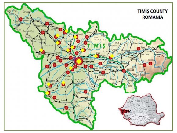
4. a) Study Area
5. b) Blood Sampling
The epidemiological study was carried out from February 2008 to October 2010, with 457 asymptomatic dogs, of different breeds, aged between 6 months and 15 years. In this group of dogs 183 were females and 274 males. Dogs examined were from 42 localities in Timi? County. Some of them belonged to different owners and some dogs were from shelters. Blood samples were collected into vacutainers with EDTA anticoagulant.
6. ( D D D D ) G
The liquid was centrifuged, and the sediment was mixed with equal parts of 1:1000 methylene blue dyes. The colored sediment was spread on a slide, and covered with coverslip, and was examined under the microscope, using the 10x and 40x light microscope objective.
7. d) Molecular Techniques
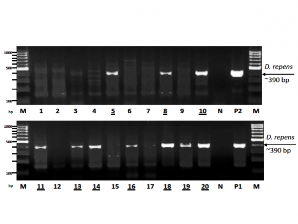
Positive blood samples with microfilariae were examined using molecular techniques, at the Department of Parasitology and Zoology of Faculty of Veterinary Science "Szent István" University in Budapest, Hungary, in 2009 and 2010. The DNA extraction was performed in 100 ?l of blood collected on EDTA, for each positive sample. We used the Blood and Tissue Dneasy kit (QIAGEN). The general primers and the thermal profile described by Casiraghi et al., 2006 [6] were used for the PCR, with species specificity for detecting D. repens and D. immitis presence. The migration of amplicons was performed by electrophoresis in 1.5% agarose gel. The ethidium bromide dye was used for preparing the agarose gel. After the migration of PCR products through the agarose gel, the gel image with the migrated DNA fragments was photographed using a Kodak EDAS 290 Polaroid system. The PCR-products were sequenced by Biomi Ltd. (Gödöll?, Hungary) directly using the ABI technology, in order to verify the specificity of PCR reactions. e) Diro Speed® Test / Heartworm (Bio Veto Test, France)
Five samples (5/33) positive for microfilariae, not examined by PCR, were investigated to detect D. immitis antigen. The examination of the samples was processed according to the manufacturers description.
8. III.
9. Results
As a result of the examination of blood samples, using the modified Knott's method, the microfilariae were identified in 33 of the 457 dogs examined (Table 1). The prevalence of Dirofilaria spp. in dogs, according to the modified Knott's method was 7.2%. The microfilariae examined using the Olympus microscope with video extension, and 400 x objectives, showed the morphology of D. repens microfilariae. This means the absence of the cephalic hook, the front end having a slightly tapered shape, and the filiform caudal end presented an "umbrella handle" shape (Figure 1,2,3). The length of the microfilariae observed was between 330-380 ?m.
The dogs infected with Dirofilaria spp. belonged to 16 breeds and 17 of them being males and 16 females aged between 2 and 13 years (Table 2, 3).
The size of the fragments amplified for the 12SrDNA in 18 samples (18/28) were ~ 390 base pairs and suggested that the samples isolated from dogs with microfilaremia belonged to the D. repens species, and ~ 450 base pairs at a sample of 28, that suggested an infection with D. immitis. An example is shown in Figure 4.
Only one dog tested positive for infection with D. immitis, a Rottweiler breed female, aged 11 years, from Timi?oara. The result of the infection with D. immitis, obtained by PCR, is questionable. At this patient further investigation were not possible. The number of microfilariae (mf) / ml of blood in dogs with dirofilariosis ranged from <30 to 5000 mf / ml of blood. This molecular biology study, performed for the first time in Romania, shows that the dogs diagnosed with microfilaremia were infected with D. repens and D. immitis. Tested with the Speed®Diro / Heartworm (Bio Veto Test, France), all 5 dogs (5/33) examined were negative for antigens of D. immitis.
10. IV.
11. Discussions
Most dog of diagnosed with microfilariae of Dirofilaria spp. came from localities crossed by a river, such as Timi?, Albina, Bega Veche, Behela rivers and the Bega Channel (Figure 5). The rivers, the lakes and the ponds are considered as preferred habitats for mosquito's larval development. So far, there are no statistics available concerning the species of mosquitoes identified in Timi? County. The increasingly high temperatures in summer, the mild winters, the large number of vectors, and numerous stray dog and the dog imports, has led to dirofilariosis spread in the Western part of Romania.
The dogs with microfilaremia were considered local cases because they have not left our country and have not been imported from Europe. Taking into consideration the results of this research we concluded that the infection with D. repens is endemic in the Western part of Romania.
The number of dogs carried subcutaneous dirofilariosis is growing, and this is why this study should be continued. The D. immitis parasitism was not identified so far in the Western part of Romania. The case tested positive by PCR in this survey, is the first one found.
Between 2008 and 2010, several necropsies in dogs, were achieved at the Necropsy Pathology Laboratory in Timi?oara, for "finding" D. immitis. Special attention was given to the thoracic cavity and heart. Using this technique, only adult D. repens were found, located in the subcutaneous tissue in certain body regions. An interesting case was represented by a dog, which presented a D. repens nematode in the scrotum after orchidectomy. This surgical procedure was performed in a veterinary clinic EUROPET-FV in the city.
In Hunedoara County (central-western part of the country) 92 dogs were tested at the shelter. Two dogs were diagnosed with D. repens (2.2%, 2/92) [7]. In 2009 -2010, in Arad, 30 dogs were tested for microfilariae. A single dog was diagnosed with subcutaneous dirofilariosis (3.3%, 1/30) (Ciocan, unpublished observations).
In Bucharest (Southeast part of the country) the infection with D. immitis and D. repens of dogs had been confirmed long time ago. Coman et al. (2007) from the "Spiru Haret" University of Veterinary in Bucharest confirms the presence of D. immitis in 12 dogs examined (23.1%, 12/52) [8]. Tudor et al. (2008) reported a prevalence of 29.3% (34/116) for the D. immitis infection in dogs in the Bucharest area [21]. A year later, in Bucharest the diagnosis of D. immitis was confirmed again, after the parasitological examination of 35 dogs [20].
In Ia?i (Northeastern part of Romania) in March 2009 was reported the first case of infestation with D. immitis in dogs, and afterwards 27 new cases were diagnosed in dogs raised by owners and 41 cases in dogs from shelters. A total number of 303 dogs were examined and 68 of them tested positive for microfilariae (Knott's method), with a prevalence of 22.4% (D. immitis). Using the Snap Heartworm rapid test, 16 dogs tested positive for D. immitis (5.3%). The first case of D. repens infestation was diagnosed in February 2009 [1,2]. Acatrinei et al. (2008) confirms the presence of D. immitis in four dogs from the southeast part of the country (Tulcea) [3].
V.
12. Conclusions
Following the research, we concluded that the prevalence of D. repens infection is high in Timi? region. The Western Region meets all the conditions for developing of Culicidae larvae and therefore canine dirofilariosis may increase in the future.
The presence of stray dogs in many places represents a reservoir in the spread of both dirofilarioses in other animals and humans. It is pointed out that Timi? County bordered with Hungary and Serbia where the subcutaneous dirofilariosis cases in dogs are very frequent. A special attention should be given on imported dogs. Veterinarians and dog owners should be informed of the presence of D. immitis and D. repens in this part of Romania and therefore, it is very important to inform the dog owners about the available prevention possibilities.
13. VI.
14. Volume XIII Issue II Version I

| Sex | n | Knott's test positive | Prevalence (%) |
| Males | 274 | 17/274 | 6.2% |
| Females | 183 | 16/183 | 8.7% |
| Total | 457 | 33/457 | 7.2% |