1. Introduction
eiomyoma arising primarily in ovary is a rare tumor and less than 60 cases have been reported till date 1 . It accounts for just 0.5 to 1 % of all benign ovarian tumors 2 . The majority of them are small, measure only a few millimeters and most (80%) occur in premenopausal age group 3 . They probably originate from smooth muscle cells in the ovarian hilar blood vessels but there are other possible origins including cells in the ovarian ligament, smooth muscle cells or multipotential cells in the ovarian stroma, undifferentiated germ cells 4 or they may arise from cortical smooth muscle metaplasia, smooth muscle metaplasia of endometriotic stroma, smooth muscle present in mature cystic teratoma and smooth muscle in walls of mucinous cystic tumor as depicted by various cases reported till now [5][6][7] . We report here a case of relatively large (2.5 x 2.0 cm) ovarian leiomyoma incidentally diagnosed in a 65 year old female with bilateral serous cystadenoma.
Author ? ? ? ? ¥: Department of Pathology Pt. B D Sharma Post Graduate Institute of Medical Sciences, Rohtak-124001, Haryana India. e-mail: [email protected] II.
2. Case Report
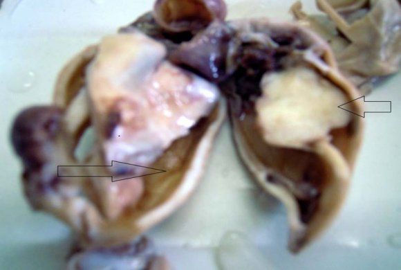
A 65 year old female presented with backache since 1 month and pain in lower abdomen since 20 days. There were no other complaints. The pelvic examination and transvaginal ultrasonography showed the presence of bilateral ovarian masses. Intraoperative frozen section revealed serous cystadenoma in both ovaries and adenofibroma like areas in left ovary. She underwent Pan hysterectomy with bilateral salpingooophorectomy. Grossly, Uterus and cervix measured 5.0 x 3.0 x 2.0 cm. Right fallopian tube measured 5.0 cm in length and lumen was dilated. Right ovary measured 3.0 x 2.0 cm and cut section showed a small cyst measuring 0.4 cm in diameter. A larger cyst measuring 5.0 x 3.5 x 2.0 cm was also found attached to the right ovary. Left fallopian tube measured 1.5 cm in length. Left ovary was replaced by a cystic structure measuring 7.0 x 4.5 x 3.5cm. An attached solid area was also identified measuring 2.5 x 2.0 cm which was encapsulated and grey white. On microscopic examination, endometrium showed changes of cystic atrophy while myometrium was unremarkable. Sections from cystic areas in both ovaries revealed serous cystadenoma . The solid areas in left ovary showed whorling of uniformly spindle shaped smooth muscle cells with eosinophilic cytoplasm and oval bland nuclei. There was negligible pleomorphism, nuclear atypia and only 1-2 mitotic figures per 10 high power fields. A possibility of benign smooth muscle tumor was considered. Special stain like Masson's trichrome showed the presence of smooth muscle. Immunohistochemistry showed positivity for ?-smooth muscle actin and desmin confirming the existence of smooth muscle. The final diagnosis of leiomyoma of the ovary with serous cystadenoma was offered.
3. III.
4. Discussion
Most ovarian leiomyomas are small, measuring only a few millimeters in diameter and are assosciated with ipsilateral or contralateral ovarian lesions [5][6][7] . But to the best of our knowledge, this is the first case of a primary ovarian leiomyoma assosciated with bilateral serous cystadenoma. Possible origin of this leiomyoma may be smooth muscle present in wall of serous cystadenoma. Hameed showed that leiomyoma of ovary can arise from smooth muscle of mucinous cystadenoma 10 .
Ovarian leiomyomas are asymptomatic and are found incidentally at surgery or at autopsy [2][3][4] . Some rare cases may be symptomatic and may present with abdominal pain, a palpable mass, hydronephrosis, elevated CA-125, hydrothorax and ascites 8 . In our case, pressure symptoms were due to bilateral serous cystadenoma rather than leiomyoma itself.
Ovarian leiomyoma is associated with its uterine counterpart in 78 % cases 2 . In our case, no uterine leiomyomas were identified even after careful serial sectioning, which makes it a primary tumor of the ovary. Primary ovarian leiomyomas are itself a rare entity and its occurrence in this postmenopausal female makes it more interesting.
Although whorling pattern and shape of smooth muscle cells of ovarian leiomyoma is quite characteristic, but, due its rarity several other tumors should be included in the differential diagnosis. Differential diagnosis of ovarian leiomyoma are fibroma, thecoma, cellular fibroma and sclerosing stromal tumor [9][10][11] . It can also be confused with tumors arising from broad ligament and extending into the hilum of ovary or wandering leiomyoma. Masson's trichrome stain helps to distinguish smooth muscle from fibrous component in the lesion. Moreover, desmin shows diffuse positivity in leiomyomas whereas fibromatous tumors are negative or only focally positive. But, ?-SMA is positive in both leiomyomas and fibromatous tumors and thus can't differentiate between the two 12 . Thecomas do not express ?-SMA and are positive for ?-inhibin and calretenin. Leiomyosarcoma, although very rare, should also be ruled out using multiple criteria like mitotic count, cytological atypia and tumor necrosis1. Treatment of ovarian leiomyoma is cystectomy or ovariectomy or ovarian wedge resection 10 .
5. IV.
6. Conclusion
To conclude, this case is a primary ovarian leiomyoma considering histopathological and immunehistochemical features. The postmenopausal patient, relatively large size (2.5 cm), absence of uterine counterpart, association with bilateral serous cystadenoma makes this case rarest of its type. Thus, despite its rarity, ovarian leiomyomas should always be considered as a possibility whenever spindle cell lesions of ovary are suspected. Appropriate diagnosis and ruling out a malignant lesion requires extensive tumor sampling and additional immunohistochemical analysis. Overall, since it is a benign tumor, ovary preserving surgery is performed in young females to preserve fertility in these women.