1. I. Introduction
Bovine tuberculosis is a contagious disease, which can affect most warm blooded animals, including human being (Radostits et al., 2007). Organisms are excreted in the exhaled air, in sputum, feaces (from both intestinal lesions and swallowed sputum from pulmonary lesions), milk, urine, vaginal and uterine discharges, and discharges from open peripheral lymph nodes of infected animals (Radostits et al., 2007). In cattle, exposure to this organism can result in a chronic disease that jeopardizes animal welfare and productivity and in some countries leads to significant economic losses by causing ill health and mortality (Ewnetu et al., 2012). Moreover, human TB of animal origin caused by M. bovis is becoming increasingly evident in developing countries (Russel, 2003;Mamo et al., 2013).
Bovine tuberculosis diseased animal loses 10 to 25% of their productive efficiency; direct losses due to the infection become evident by decrease in 10 to 18% milk and 15% reduction in meat production (Radostits et al., 1994). Apart from effects on animal production, it has also a significant public health importance (Müller et al., 2013). Currently, the disease in human is becoming increasingly important in developing countries, as humans and animals are sharing the same micro environment and dwelling premises, especially in rural areas, and susceptibility of AIDS patients to tuberculosis (Shitaye et al., 2007) (Smith et al., 2006;Pal, 2007;Malamaet al., 2013). Although, recent studies indicated that M. tuberculosis has been isolated from cattle (Ameni et al., 2011) and M. bovis from humans infected with bovine tuberculosis (Zeweld, 2014), M. tuberculosis is specifically adapted to humans while M. bovis is most frequently isolated from domesticated cattle (Smith et al., 2006), In spite of variation in host specificity, the members of MTBC are characterized by 99.9% or greater similarity at nucleotide level and are virtually identical at 16s rRNA sequence (Brosch et al., 2002). T causes 10 to 15% human cases of tuberculosis in countries where pasteurization of milk is rare and bovine tuberculosis is common (Ashford et al., 2001;Berg et al., 2015).
In developing countries like Ethiopia, the socio economic situation and low standard living area for both animals and humans are more contributing in TB transmission between human to human and human to cattle or vice versa (Ameni et al., 2010a;Ejeh et al., 2013). Human infection due to M. bovis is thought to be mainly through drinking of contaminated or unpasteurized raw milk and under cooked meat. The high prevalence of TB in cattle, close contact of cattle and humans, the habit of raw milk and meat consumption, and the increasing prevalence of HIV may all increase the potential for transmission of M. bovis and other Mycobacteria between cattle and humans (Shitaye et al. 2007).
Bovine tuberculosis is an endemic disease of cattle in Ethiopia, with a reported prevalence of 3.5-5.2 % in abattoir (mostly zebu) and 3.5-50% in crossbreed farms (Shitaye et al., 2007;Demelash et al. 2009;Regassa et al., 2010;Berg et al., 2011). Nevertheless, the available information is limited due to inadequate disease surveillance and lack of better diagnostic facilities (Cosiviet al., 1998;Asseged et al., 2000). In particular, information on genotypic characteristics of M. bovis, a strain affecting the cattle population in Ethiopia, is limited (Biffa et al., 2010a). Such information is critical to monitor transmission and spread of the disease among cattle (Berg et al., 2011).
The World Health Organization 2009 report indicated that the status of TB in Gambella Region was the highest from all the Ethiopian Regions, with the notification rate (new and relapse) 261-421/100, 000 (WHO, 2009). This was one of the bases of the present study.
Gambella regional state has large livestock populations. Despite, the large number of livestock population in the region, there is no information on BTB. Despite the fact that bovine tuberculosis is a public health threat and also leads to economic losses, in Ethiopia research on and control of animal tuberculosis has not received much attention like human tuberculosis (Chukwuet al., 2013).Thus the present study was designed to determine the prevalence of bovine tuberculosis in Gambella town municipal abattoir and identifying risk factors associated with bovine tuberculosis, to isolate and molecular characterization of Mycobacterial isolates from slaughtered cattle and from human pulmonary TB patients and to investigate the potential risk factors for zoonotic transmission of mycobacterial infections.
2. II. Materials and Methods
3. a) Study Area
The study was conducted in Gambella town municipal abattoir and Gambella hospital of Gambella regional state, southwest Ethiopia from December 2014 to May 2015. The Gambella People's Regional State is located south west Ethiopia between the geographical coordinates of 6 0 28'38" to 8 0 34' North Latitude and 33 0 to 35 0 The Gambella town municipal abattoir: -The abattoir which is administered under Gambella town municipality is the only source of inspected beef for all inhabitants of the town. The average number of animals slaughtered per day during the study period was about 25 with all 100 % of the slaughtered animals being cattle. The overall abattoir sanitary environment is below the requirements of good hygiene practices (GHP) in slaughterhouses. The internal and external facilities and sanitary conditions of the slaughter house were very poor. Neither place for disposal of condemned carcasses nor facilities for wastewater treatment exist and it is not friendly with the environment. The abattoir workers had no clothing, boot, apron and other accessories. Three assistant meat inspectors were delivered services only during ante mortem and no one was carried out post mortem examination during the study period in such a ways the population is endanger of meat born zoonosis and sanitation problems.
4. b) Study Population and Study Design
According to the available logistics and time a total of 500 apparently healthy animals slaughtered in the abattoir of Local Nuer, Horro and Felata breed cattle were included as study population for the stated objectives and the major sources of cattle for this abattoir were Gambella town and its surroundings, Mettu, Gore, Bure, Sibo and Gumero. In addition, 50 Acid fast bacilli (AFB) positive sputum samples from human TB patients attending the health facilities in Gambella town were included.
A cross sectional study with systematic random sampling was carried out in abattoir to examine the carcass and sample suspected TB lesions from slaughtered cattle at Gambella town municipal abattoir. Similar cross sectional study and purposive sampling was carried out to collect samples from all AFB positive TB patients attending Gambella Hospital. Both sputum and extra pulmonary TB samples mainly fine needle aspirate from suspected human case was taken in the course of the study period for isolation and molecular characterization of the causative agents.
5. c) Sampling, Sample Size Determination and Study
Methodologies All animals coming to the slaughter house from different areas during the study period were considered for sampling. The sample size calculation was based on 50% prevalence assumption (since there was no study on bovine tuberculosis in the area), 95% CI and d=0.05 (Thrusfeild, 2005).
n = Z 2 x p expe (1-p expe ) d 2Where n= required sample size P exp . =expected prevalence d=Desired absolute precision (5 %) Z= Normal distribution constant Therefore, the sample size calculated was 384, but to increase the precision using thumb rule by 20% and the total animals supervised were 500.
The sample size for the questionnaire survey used was 100 for livestock owners, and abattoir workers. For human case, a total of 50 acid fast positive patients were interviewed about their association with cattle, habit of consumption of meat and milk and other relevant information related to tuberculosis.
6. d) Ante and postmortem examination
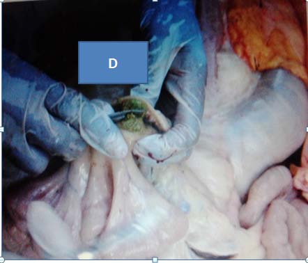
Physical examination of the animals were carried out before they were slaughtered. Body temperature, pulse rate, respiratory rate, condition of superficial lymph nodes and visible mucus membranes were examined and recorded for individual animals to be slaughtered. Breed, source or origin and sex were also recorded. Age was estimated as described by Amstutz (1998) and Body Condition Scoring (BCS) chart was made based on the description by Nicholson and Butterworth (1986). Detailed postmortem examination (inspection, palpation and incision) of the carcass, lungs, liver and kidneys together with mesenteric, hepatic lymph nodes and lymph nodes of the head was undertaken in accordance with the method developed by Ethiopian meat inspection and quarantine division of the Ministry of Agriculture (Hailemariam,1975;Ameniet al., 2007). Lymph nodes were incised into a size of 2 mm to facilitate the detection of tuberculous lesions from each animal. These include Mandibular, Retropharyngeal, Bronchial, Mediastinal, and Mesenteric lymph nodes. The animal was classified as lesioned (infected) when tuberculous lesion was found, and if not as non lesioned (not infected). The severity of gross lesions in individual lymph nodes and other organs were scored as follows; 0= no gross lesion, 1= small lesion at one focus, 2= small lesions at more than one focus and 3= extensive necrosis as developed by Ameni et al. (2006). The cut surfaces were examined under bright light for the presence of abscess, cheesy mass, and tubercles (Corner et al., 1990). In the presence of suspected tuberculous lesion, tissue samples were collected in sterile universal bottles containing 0.85% normal saline for culture kept at -20 0 crefrigerator. The samples were transported under cold chain by ice box with packed ice to the Akililu Lema Institute of Pathobiology for culture and further processing in three week basis.
7. e) Isolation of mycobacteria
Tissues with suspected lesions were collected and subjected to bacteriological culture examination. The tissue specimen or sputum collected from AFB and gene Xpert positive patients for culture were collected individually in to sterile universal bottles in normal saline and then labeled and kept frozen ( ?20 °C) at Gambella regional hospital before being transported to Aklilu Lema institute of Pathobiology laboratory Addis Ababa.
The specimens were labeled and pooled together, kept in universal bottle containers, and then transported in ice pack box to Aklilu Lemma Institute of Pathobiology, Addis Ababa Ethiopia, within three week basis by airplane. There the samples were processed for isolation of M. tuberculosis complex according to the standard methods .
8. f) Identification and characterization of mycobacteria
Initial identification of mycobacterial species from animal tissue was based on the rate of growth, pigment production, and colony morphology as described in OIE (2009). When visible colonies were observed, Ziehl Neelsen staining was performed to confirm the presence of acid-fast bacilli. AFB positive isolates were prepared by mixing two loops full of colonies in 200 mL distilled water, heat-killed at 80 0 C for 1 hour using water bath, and stored at -20 0 C until molecular characterization was perform and were subjected to PCR based on amplification of a multi copy DNA target sequence for identification of M. bovis and M. tuberculosis (Debebe et al., 2013).
9. g) RD deletion typing
For RD9 deletion typing of culture positives of sputum; RD9 intR: CTG GAC CTC GAT GAC CAC TC, RD9 flankF: GTG TAG GTC AGC CCC ATCC and RD9 flankR: GCC CAA CAG CTC GAC ATC primers to check for the presence of RD9 locus was used; The HotStarTaq Master Mix system from Qiagen was used for PCR, with primers described previously (Ameni et al., 2013).
The primers used were RD4Flank int: 5´ACAC GCTGGCGAAGTATAGC3´; RD4flankR: 5´AAGGCGA ACAGATTCAGAT3´ and RD4falnkF: 5´CTCGTCGAAG GCCACTA AG3´. The mixture was heated in a Thermal Cycler (Applied Bio-systems; Gene AMP 9700) for 15 minutes at 95°C and then subjected to 35 cycles of one minute duration at 95°C, one minute at 55°C, one minute at 72°C and 10 minutes at 72°C. The presence of RD4 (RD4 is intact in M. tuberculosis, M. africanum) gives a product size of 335 bp (RD4 intF + RD4flankR), and its absence (M. bovis) gives a product size of 446 bp (RD4flankF + RD4flankR).
10. h) Spoligotyping
Spoligotyping was carried out using the commercially available kit according to the manufacturer's instructions and as previously described by Kamerbeek et al. (1997).
11. i) Questionnaire survey
The roles of various risk factors in the occurrence and spread of bovine TB among cattle, and between cattle and people, were assessed by a questionnaire. Structured questionnaire was distributed to TB patients, cattle owners, and abattoir workers to assess the perception of stakeholders on the occurrence of bovine tuberculosis, livestock constraints, socioeconomic status, herd composition, awareness on the potential risk of zoonotic transmission of bovine tuberculosis.
12. j) Data Management and Analysis
Prevalence was calculated as the proportion of suspected lesion positive animals from the total number of animals visited (Thrusfield, 2005). Data related with age, sex, breed, origin and body condition of each animal was recorded on a data sheet during antemortem examination. Presence or absence of TB like lesions and affected tissues were recorded during postmortem examination. The recorded data was entered and stored in Microsoft Excel computer program and analyzed by STATA version 11 (STATA Corp. College station, TX). The variations between different factors were also analyzed using multi variable logistic regression and chi-square (?2) was used for association of different risk factors. A p-value <0.05 was considered statistically significant, 95% confidence interval was considered and Odds ratio analysis was used.
In molecular epidemiology study of isolates from human pulmonary tuberculosis patients and animals tissue, the spoligotype patterns were converted in to binary and octal formats and entered to the online spoligotypedatabase, http://www.pasteur guadel oupe.fr:8081/SITVIT Demo/index.jsp to determine the shared international spoligotype (SIT) number and the results were compared with already existing designations in the international spoligotyping database (SpolDB4.0 database). Those isolates with no designated SIT number were considered as new to the database. Two or more isolates with identical spoligotype pattern were considered as clustered while those with single SIT were considered as non-clustered isolates. TB-lineage and family were determined using SPOTCLUST database, http://tbinsight.cs.rpi.edu/about _spotclust.html
13. k) Ethical Considerations
Ethical clearance was obtained from the Ethical Committee of Gambella regional health office (Ref. number of 16/3776/7) and working permission was gotten from the hospital higher managers and the municipality.
14. III. Results
15. a) Prevalence of Bovine Tuberculosis
The overall prevalence of bovine tuberculosis in slaughtered cattle of Gambella municipal abattoir was 13.2% (66/500: 95% CI, 10.22-16.18) based on the occurrence of gross tuberculous lesions.
16. *Statistically significant b) Gross Pathology Results
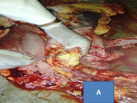
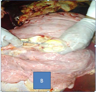
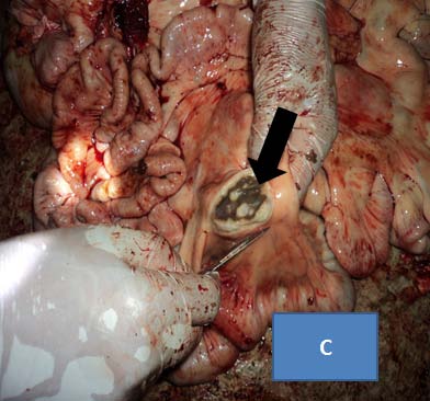
Gross lesions were observed in the lymph nodes and lung of the slaughtered cattle and the majority of the lesions were considered typical of tuberculous lesions characterized by central round, oval, or irregular, often coalescing areas of caseous necrosis and mineralization (calcification) (Figure 1). Whenever gross lesions suggestive of TB were detected in any of the tissue, the tissue was classified as having lesions. The frequency and distribution of lesions according to organ level and anatomical site is indicated in (Table 2).
17. c) Mycobacteriological Culture Result
Out of 82 tissue samples 14(17.07%) showed a growth on LJ medium and out of 50 sputum samples and one FNA sample, 17(34%) of sputum samples had showed growth on LJ media while the FNA sample did not grow (Table 4).
18. IV. Discussion
Tuberculosis remains a major global health problem causing high morbidity and mortality among millions of people each year (WHO, 2014). Tuberculosis caused by M. bovis is clinically indistinguishable from tuberculosis caused by M. tuberculosis and globally the proportion of human tuberculosis caused by M. bovis is estimated to 3.1% of all forms of which 2.1% of pulmonary and 9.4% of extra pulmonary forms (Cosivi et al., 1998).
Ethiopia is one of the countries with highest number of livestock resource in Africa and animal tuberculosis is known to be endemic and wide spread in the country. However, in spite of high prevalence both human and animal tuberculosis in the country, the emphasis given on bovine tuberculosis to the Gambella region is very little and so far no research were carried out on BTB in Gambella Region. Infection of cattle with M. bovis constitutes a human health hazard as well as an animal welfare problem. Furthermore, the economic implications in terms of trade restrictions and productivity losses have direct and indirect implications for human health and the food supply (Zeweld, 2014).
In the present study an attempt was made to determine the prevalence of bovine tuberculosis in Gambella town municipal abattoir and identifying risk factors associated with bovine tuberculosis, to isolate and molecular characterization of Mycobacterial isolates from slaughtered cattle and from human TB patients and to investigate the potential risk factors for zoonotic transmission of mycobacterial infections from animal to human and vice versa.
Based on detailed post mortem inspection the prevalence of BTB in slaughtered cattle was found to be 13.2%, which is moderately high and this result was comparable with other pervious research reports carried out on cattle originated from extensive and pastoral production system of Ethiopia; 11.50% by Abdurohaman (2009) in Butajira, and 11% by Mamo et al. (2013) in Afar, but less than a result from 19.8% record from cattle slaughter in rural Tanzania (Cleaveland et al., 2007). The result of the present prevalence study was higher than findings by various other authors Biffa et al., 2010a who reported 4.2% prevalence in cattle slaughtered at in Yabello municipal abattoir and 4.5% at Hosaana abattoir by Teklu et al., (2004). In addition, the result were also higher than previously reported by other researcher in Northern and cental parts of the country ( ). This difference in prevalence of tuberculous lesions could be due to the difference in origin or types of production system and breed of animals that are slaughtered in the abattoirs.
In respective of small sample size due to wondering of the Felata breed from place to place, association of breed with prevalence of BTB showed a statistically high significant difference among different local breed of cattle, (P = 0.000) animals which might be related to the genetic difference of the breeds, Other previous studies also showed different breeds could result in difference in susceptibility to BTB infections .
There is a statistically significant difference in the prevalence of the disease (P = 0.000) between BCS, the prevalence being the highest in poor body condition (32.6 %) as compared to medium (12.7% ) fatty (good) animals (6.3 %) respectively which in agreement with study resulted by Nemomsa (2014). This could due to related to the weak protective immune response in poor body conditioned animals as compared to good one that may result extensive lesions and wasting of the body condition as well as its chronicity nature of the disease. The present result is consistent with previous reports which indicated that animals with good BCS have relatively good immunological response to the infectious agent than animals with medium BCS (Radostits et al., 1994;Radostatit et al 2007).
In this study, gross tuberculosis lesions were found most frequently in the lymph nodes of thoracic cavity (50%), mesenteric lymph node (25.6%), followed by lymph nodes of the head (24.4%).This finding is significantly different from previous studies done in Ethiopia (Tamiru et al., 2013) where 70 and 70.7% TB lesions were reported in lungs and associated lymph nodes, respectively. However, the distribution of TB lesion in the current study significantly similar with reports from Mexico (Ndukum et al., 2010) where 49.2% of lesions involved the thoracic lymph node. The result, therefore, indicate that the primary route of infection was through the respiratory route which can also spread to other parts of the body as described previously (Radostits et al., 2007).
In this study, culture positivity in primary culture media was found low and confirmed in 23.49% (31/133) despite slightly lower than that reported by , 56% culture positivity. This low isolation rate of mycobacteria may have resulted from reduced sensitivity of culture arising from prolonged storage at field sites and the freeze-thaw cycles that occurred during transportation and contamination of tissue samples (OIE, 2009). Furthermore, the presence of caseous and/or calcified lesions and even lesions resembling tuberculous lesions may not always found to be of mycobacterial origin; they can be caused by any other intracellular organisms or parasites, or viable mycobacteria may not be present in calcified lesions (Corner, 1994).
In the present study, interestingly, M. tuberculosis strain SIT523 was isolated and characterized with spoligotyping from cattle cranial Mediastinal lymph node tissue and the result implies the occurrence of reverse zoonosis in the study area where human strains could be transmitted to cattle. The transmission to cattle could be through different routes including ingestion of feed contaminated with infected sputum and/or urine from M. tuberculosis infected farmers. Humans suffering from active TB are the most probable source of M. tuberculosis in animals, with infection spread via sputum, and rarely urine or faeces (Thoen and Steele, 1995) or respiratory route as in rural area of Ethiopia, grazing cattle are commonly brought into the farmers' households at night where they may become infected via aerosol transmission from humans (Ameni et al., 2013). Previous studies in Ethiopia had confirmed transmission of M. tuberculosis from farmers to their cattle, goat and camel (Berg et al., 2009 (Tsegaye et al., 2010). On to this, the identification of M. tuberculosis from cattle tissues requires further investigation.
In molecular characterization of isolates from human tuberculosis patients, M. tuberculosis was the predominant species causing TB in human and the genetic diversity of the isolate on the spoligopattern was 45.45%, which was higher than previous reports in other part of Ethiopia where 39% of spoligotype based genetic diversity where reported in Afar PTB patients (Mamo et al., 2013). The difference might be related to difference on geographic and sociocultural difference among the studied population which might affect the transmission pattern of the organism. The most common spoligotype identified from TB patient was the SIT 289, in agreement with previous study (Ameni et al., 2011) which also reported the same SIT289 strains in pulmonary patients of central Ethiopia. In the present study, the predominant lineage was unknown according to TB-insight database analysis. Similar, unknown lineage had been previously reported form patients from Northwestern Ethiopia (Belay et al., 2014) and this indicates the need for further investigation.
In the present study, the questionnaire survey of the respondents showed that 22 % of them were aware of BTB with no knowledge about zoonosis of the disease. This disagrees with report from Tamiru et al., (2013) 80.7% of them were aware of BTB with low level knowledge about zoonoses of the disease. This result was comparable with the study on assessment of the knowledge of cattle owners about BTB in WuchaleJida district, Ethiopia showed that 38.3% (36 of 94) of the respondents knew that cattle can have tuberculosis, and 30.8% (29 of 94) recognized that BTB is zoonotic (Ameni et al., 2003). have indicated that lack of understanding regarding the zoonotic of BTB, food consumption behavior and poor sanitary measures is the potential risk of BTB to public health. The proportion of BTB contributes to total tuberculosis cases in humans depends on the prevalence of the disease in cattle, consumer habits, socio-economic conditions, level of food hygiene (Ashford et al., 2001) and medical prophylaxis measures in practice (Tigre et al., 2011). According to the result of the present study, 45% consume unpasteurized or raw milk. Similarly, studies conducted in different parts of Ethiopia indicated the habits of raw milk consumption. The current result on habit of milk consumption was lower than 85.7% report from Jimma town, Ethiopia (Tigre et al., 2011). Study conducted in WuchaleJida district indicated 52.1% (49 of 94) households' has habit of consuming raw milk (Ameni et al., 2003), which is significant when compared with the current result. No one of the respondents in our study were found to be aware about the transmission of the disease from cattle to human and vice versa.
In our study, keeping cattle in close proximity to their house and calves in their house was a common practice of households. This indeed can facilitate transmission of the causative agent from animal to human or vice versa. According to Bogale (1999), conditions such as customs of consuming raw milk, keeping cattle in close proximity to the owner house and using cow dung for plastering wall or floor and as source of energy for cooking do exacerbate the chance of spread of tuberculosis as zoonosis in Ethiopia.
19. V. Conclusions and Recommendations
The result of the present study has shown that bovine tuberculosis was prevalent in cattle slaughtered at Gambella municipal Abattoir with moderately high prevalence (13.2%). This study also revealed that a high proportion of tuberculous lesion in the thoracic cavity lymph nodes and which implies that respiratory route was the major means of transmission. Isolation and molecular characterization of one M. tuberculosis isolate (SIT523 strain) from animal tissue sample suggested the occurrence of transmission of the agent between the communities and animals that implies reverse zoonosis. The high genetic diversity (45.5%) of the human M. tuberculosis isolates (SIT289, SIT134, SIT1634, SIT142, and the new one) and presence of clustering of the isolates might indicate the recent transmission pattern and circulation of the agents in the study communities. Lack of awareness regarding BTB and its routes of transmission in the study population was high and existence of habits of consumption of raw animal product and sharing of the same microenvironment with their livestock could be potential risk factors for zoonotic transmission of the disease. On the basis of findings of the present study, the following recommendations are forwarded: Further study should be conducted with larger sample size and geographic coverage to elucidate the role of M. tuberculosis complex in human and animal, With the finding of promising result on molecular characterization using few samples; a broader study to investigate the molecular epidemiology in human and animal tuberculosis is essential, Public health awareness campaigns should be launched and needed to raise community awareness about the risk of BTB transmission through consumption of raw/under cooked meat; and the zoonotic implication of BTB/, route of reverse zoonosis are of extreme importance for effective implementation of TB control measures, Establishment of collaboration between physician and veterinarians to trace back positive patient to get profile of their cattle in the slaughterhouse across


| Year 2015 | ||
| uberculosis is | communicable | Mycobacterial |
| disease of human and animals, caused by | ||
| members of Mycobacterium tuberculosis complex | ||
| (MTBC) | ||
| Research ( ) F | ||
| Global Journal of Medical | ||
| © 2015 Global Journals Inc. (US) | ||
| lower altitudes varies from 900-1,500mm; at higher |
| altitudes it ranges from 1,900-2,100mm. The annual |
| evapotranspiration in the Gambella reaches about |
| 1,612mm and the maximum value occurs in March and |
| is about 212 mm (Tilahune, 2012). Based on the 2013/ |
| 2014 Census conducted by the Central Statistical |
| Agency of Ethiopia (CSA), the Gambela Region has total |
| population estimation of 406,000 (CSA, 2013/2014) and |
| livestock population of Gambella 253,389 cattle, 39,564 |
| sheep and 83,897 goat (CSA, 2010/2011). |
| Risk factor | Number examined Number positive | Crude odds ratio | Adjusted odds ratio | |
| (95% CI) | (95%CI) | |||
| Age (year) | ||||
| <5 | 24 | 2 | 1 | 1 |
| 5-8 | 188 | 24 | 1.61(0.36-7.28) | 1.26(0.25-6.43) |
| >8 | 288 | 40 | 1.77(0.40-7.84) | 1.08(0.22-5.37) |
| Sex | ||||
| Male | 346 | 36 | 1 | 1 |
| Female | 154 | 30 | 2.08(1.23-3.53) | 1.05(0.52-2.15) |
| BCS | ||||
| Anatomical site | Organ affected | Frequency (%) |
| Head | Mandibular lymph node | 5(6.1%) |
| Retropharyngeal lymph node | 15(18.5%) | |
| Thoracic | Bronchial lymph node | 8(9.8%) |
| Mediastinal lymph node | 19(23.2%) | |
| Lung | 14(17.1%) | |
| Abdominal | Mesenteric lymph nodes | 21(25.6%) |
| Total | 82(100%) |
| Tissue | Number examined | Number positive (%) | Mean ±SE) |
| Lung | 500 | 14(2.8) | 1.86±0.231 |
| Mandibular | 500 | 5(1) | 2.6±0.245 |
| Bronchial | 500 | 8(1.6) | 2.25±0.366 |
| Mediastinal | 500 | 19(3.8) | 1.5±0.159 |
| Retropharyngeal | 500 | 14(2.8) | 1.93±0.245 |
| Mesenteric | 500 | 22(4.4) | 2.14±0.168 |
| Type of Specimen | Number of | Growth on LJ- | Growth on LJ- | Growth | Total growth (%) |
| sample | pyruvate | glycerol | on both | ||
| Sputum | 50 | 8 | 9 | 5 | 17(34) |
| FNA | 1 | - | - | 0 | |
| Animal tissue | 82 | 5 | 10 | 1 | 14(17.07) |
| Total | 133 | 13 | 19 | 6 | 31(23.3%) |
| Specimen | Spoligotype | Octal number | Lineage | |
| 1 | Sputum | 1110000111111111111111000000000000111011111 | 703777740003571 | Unknown |
| 2 | Sputum | 1111111111111111111111111111110100001100111 | 777777777720631 | Euro-American |
| 3 | Sputum | 11110000111111111111111000000000000111011111 | 703777740003571 | Unknown |
| 4 | Sputum | 1111111111111111111111111111110100111111111 | 777777777723771 | Indo-Oceanic |
| 5 | Sputum | 1111111111111111111111111111110100001100111 | 777777777720631 | Euro-American |
| 6 | Sputum | 1110000111111111111111000000000000111011111 | 703777740003571 | Unknown |
| 7 | Sputum | 1010000111111011111111000000000000111011111 | 503757740003571 | Unknown |
| 8 | Sputum | 1110000111111111111111000000000000111011111 | 703777740003571 | Unknown |
| 9 | Sputum | 1110000111111111111110000000000000111111111 | 703777700003771 | Unknown |
| 10 | Sputum | 1110000111111111111111000000000000111011111 | 703777740003571 | Unknown |
| 11 | Sputum | 1111111111111111111111111111110100001100111 | 777777777720631 | Euro-American |
| 12 | Animal Tissue | 1111111111111111111111111111111111111111111 | 777777777777771 | Indo-Oceanic |
| e) BTB Awareness and risk factor Assessment | ||||
| knowledge examined in questionnaire | Responders out of 100 (%) |
| Had noticed respiratory problems in their cattle | 30(30%) |
| Aware of bovine tuberculosis (TB) | 22 (22%) |
| Know that cattle transmit bovine TB to humans | 15 (15%) |
| Know that humans transmit TB to cattle or vice versa | 0 (0%) |
| Know that milk is a source of infection | 23(23%) |
| Know that meat is a source of infection | 17(17%) |
| Drink raw milk | 37(37%) |
| Eat raw meat | 45(45%) |
| Use the same watering point with animals | 48(48%) |
| Share the same house with animals | 30(30%) |