1. I. Introduction
he medical image has seen an important development in the last two decades such as computed tomography (CT), magnetic resonance image (MRI), positron emission tomography (PET), and single photon emission computed tomography (SPECT). Now, the doctors need information from more than one modality for efficient disease detection and diagnosis. Medical image fusion is the technique of merging images belonging to different modality into a single resultant image that is provide complementary information for better analysis.
Image fusion can be grouped into three categories: Pixel level, Region level and Decision level. There are two approaches for pixel level method: spatial domain and transform domain [4]. Spatial domain fusion techniques are simple and fused image can be obtained by directly applying fusion rules on pixel values of source images such as Simple averaging, PCA (Principal Component Analysis) [5] and linear fusion [6]. But major disadvantage of spatial domain techniques are that they introduce spatial distortions in the fused image [7] and do not provide any spectral information. Transform domain techniques overcome the disadvantages of spatial domain fusion. Pyramid and wavelet transforms based techniques are the mostly used transform domain image fusion methods. Laplacian pyramid [8], contrast pyramid [9]. These methods overcome the disadvantages of spatial domain Author ? ? ?: Biomedical Laboratory, Department of electrics and Electronics, Technology Faculty. University of Tlemcen 13000, Algeria. e-mails: [email protected], [email protected], [email protected] techniques but suffer from blocking effect [7]. As a result wavelet transform based fusion [11,12] approaches are used and it was found that no blocking effect occurred during fusion process. Discrete wavelet transform (DWT) [3] and integer wavelet transform [2] preserves different frequency information but may produce specularities along the edges and shift sensitivity, poor directionality and lack of phase information [15,16]. The third type is new multiresolution methods are proposed, such as image fusion based on nonsubsampled contourlet transform (NSCT) [17]. Due to these limitations of real valued DWT. Another approach uses biorthogonal wavelet CDF 9/7 [18] based on lifting scheme provides better representation of fused image.
The objective of this research is to introduce CDF 9/7 wavelet based on lifting scheme to fuse more properties (edge, information) of different modalities such as MRI-T1 and MRI-T2 brain images (normal and tumor). The proposed algorithm is also uses the specified fusion rules of low and high frequency subbands. Experimental results on the Brain Web database show the usefulness of this wavelet family and clearly indicate its potential in medical image fusion. The rest of paper organization of this is as follows, the section 2 we present the proposed fusion algorithm steps with fusion rules. The experimental results are shown in section 3 and we compare results obtained with the existing techniques. Finally the main conclusions are summarized in Section 4.
2. II. Proposed Fusion Algorithm
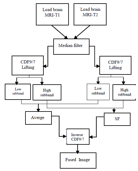
The input Images can suffer from artifacts due to different factors. However the recent advances in acquisition protocol make it possible to acquire images with very limited artifacts. The proposed method is illustrated in Figure 1 T MRI-T1 and T2 Image Fusion for Brain Image using CDF Wavelet based on Lifting Scheme a) Image de-noising using median filter (preprocessing)
This proposed system describes the information of enhancement using weighted median filter for removing high frequency component which characterize the noise. These filters have the robustness and edge preserving capabilities with noise attenuation characteristics. After the pre-processing operations the input images are subjected to wavelet analysis which is described in the following section.
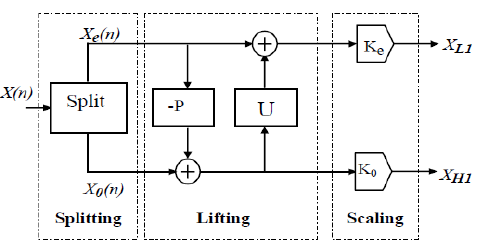
b) Biorthogonal CDF9/7 wavelet based on lifting scheme [1] Sweldens from Bell Labs proposed a method which does not depend on the Fourier transform of ascension wavelet construction in the mid 1990's presented in [19,20], where sweldens showed that the convolution based biorthogonal Wavelet Transform (WT) can be implemented in a lifting-based scheme. The lifting-based WT consists of splitting, lifting, and scaling modules and the WT is treated as prediction-error decomposition. It provides a complete spatial interpretation of WT.
In this article we deals with biorthogonal wavelet 9/7, these wavelets are part of the family of symmetric biorthogonal wavelet CDF. The low pass filters associated with wavelet 9/7 have p=9 coefficients in the analysis, p=7 coefficients to synthesize. The wavelets 9/7 have a great number of null moments for a relatively short support. They are more symmetrical and very close to orthogonality [21]. Were the first to show the superiority of the biorthogonal wavelet transform 9/7 for the decorrelation of natural images The input medical image (A, B) are decomposed using CDF9/7 lifting wavelet transform to obtain the low frequency (???? ?? , ???? ?? ) and high frequency (???? ?? , ???? ?? , ???? ?? , ???? ?? , ???? ?? , ???? ?? ) coefficients.
i
3. . Lowpass subband fusion
The approximation coefficients are averaged, to preserve the features from both the images
???? ?? = ????????(???? ?? , ???? ?? ) (1)Where ?????? is the approximation band of the fused image.
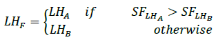
ii. Highpass subband fusion For each detailed sub bands, the spatial frequency is calculated using Equation SF.
(2)
Apply the same technique for: ???? ?? , ???? ?? , ???? ?? , ???? ?? Finally, the inverse lifting transform based on CDF9/7 wavelet is applied to generate the fused medical image.
4. III. Results and Discussion
In this section to evaluate the performance of the proposed algorithm. We fuse normal brain MRI-T1 image with MRI-T2 image from [24] data base. Similarly Brain tumor hypo intense T1 image with hyper intense T2 image are downloaded from [25].
To show the effectiveness of the multi scale transform the comparisons start with PCA (Principal Component Analysis) method, CDF9/7 DWT (discret wavelet transform) based filter banc and CDF9/7 LWT (Lifting wavelet transform) with the first level. To quantitatively compare the performance with existing fusion algorithms, entropy EN, mutual information MI, spatial frequency SF, standard deviation SD and time calcul T , are defined and calculated in the following section. These images were tested on Intel Core (I3) 2.13 GHz PC with 2GB of RAM using Matlab 2010a.
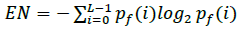
5. a) Entropy
Entropy is the quantity of information contained in a series of events. Entropy is a criterion that measures the level of information in the image, more entropy is large, more diffuse the image information.
(3)
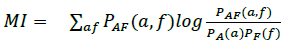
6. b) Mutual Information (MI)
Compares the image source and the fused image, more value is small, the relationship between the two images is non-existent.
7. c) Standard Deviation (SD)
Is the square root of the variance, the variance of an image reflects the degree of dispersion between the grayscale values and the average value of gray levels, more the standard deviation is large, more there are dispersion (5)
8. d) Spatial Frequency (SF)
It measures the total activity and the level of clarity of an image, an important value mean that the result of fusion is good. (6) Where RF and CF are the row frequency and the column frequency respectively.
The fused image output based on proposed method illustrate in Figure 3 and 4. This visual representation is not sufficient for analysis of the obtained fusion results. Therefore we have compared the proposed method with other ones on quantitative measures which are entropy, standard deviation, spatial frequency, mutual information. For Table1 and 2, The graphical representation of all performance evaluation parameters are showns in figure 5.
9. Global Journal of