1. I. Introduction
rowth is critical feature of a children life which differentiates them from adults and it is identified as an indicator of wellbeing. The growth factor is a decisive variable in orthodontic treatment. 1 It believe one of the main uncertain dissimilarity in nature and plays an important role in etiology of malocclusion in addition to evaluation of diagnosis, treatment planning, retention and stability of orthodontically treated cases. 2 Every person have special internal clock in maturity, the facial bones growth time and the periods of accelerated or intense physiologic growth must be individualized for superior exploit bone remodeling in accurate skeletal discrepancies. 3 In orthodontics and dentofacial orthopedics, the initiation of treatment is becoming increasingly evident, as the selection of the specific treatment procedures. 4 Skeletal age estimation is vital in planning of orthodontic treatment, due to variations on timing, duration and velocity of growth. 1 Estimation of growth potential requires the assessment of the developmental age of the individual patient. A number of developmental indicators can be utilized to assess maturity; increase in body height, skeletal maturation of the hand and wrist, cervical vertebral maturation, dental development and eruption, and menarche or voice changes. 3,[5][6][7] Chronological age is measured from the birth day, which used to recognize the developmental stage of individuals, although, it is the most easily determined parameter of all the developmental ages, it is a weak growth predictor. Physiological age considers more reliable to evaluate the maturation state because each child has his or her own characteristic time clock. . 8 The onset of chronological age varies with gender, generation, population and environment, and diverges greatly among individuals. 9 Dental age is an important matter in orthodontic diagnosis and treatment planning. It is estimate by teeth development either by calcification of crowns and roots or eruption of the crowns on the radiographs. 10 Tooth formation is widely used as a growth indicator for assessment and comparison between individuals and populations in dentistry, pediatrics and anthropology, moreover forensic sciences. 11 Skeletal maturation refers to the level of development of ossification in bone. Size and maturation can vary autonomously of each other. Skeletal maturation is well identified and frequently used for measurement of biological maturity, which resolute by radiographic assessment of one or more areas in the body. 12 Hand-wrist radiographs have been used to assess the skeletal maturity stages, it is recently been inquiry. Recently the cervical vertebra maturation (CVM) method was introduced for growth assessment; allocate skeletal age evaluation and diminish the need for additional radiographic exposure. [12][13][14] Chronological age, dental and skeletal development were regarded as regular maturational indexes of developmental maturation. 1,15 The divergence between dental, skeletal and chronological age is of enormous attention in compared with ordinary growth. 12, chronological age among different populations, to our knowledge no such study are available in Sudan, therefore the present study had been designed to provide reliable age estimation for the Sudanese population and the outcome results will be of great helps for diagnosis and treatment planning for orthodontic and pediatric patients as well as to forensic dentistry.
2. II. Materials and Methods
The material consisted of the clinical files and panoramic and lateral cephalometric radiographs of 112 Sudanese children (65 males and 46 females), aged from 7 to 16 years, who attending orthodontic treatment at the clinic of Orthodontic Department, Faculty of Dentistry, University of Khartoum, between 2009 and 2015. At the begining of the study, the chronological ages of the patients were recognized from the child's birth to the day the panoramic radiograph was taken. The chronological age was recorded in years and months.
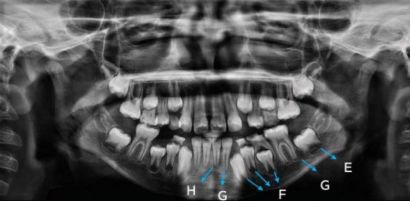
To identify the dental age and skeletal maturity stages, all OPGs and lateral cephalometric radiographs were evaluated in dark room on a view box to ensure the maximum contrast. The dental age was assessed on panoramic radiograph by using Demerjian method. 10 This method based on the developmental stages of the left mandibular permanent teeth, were rated on an 8stages scale from A to H. (figure1) The maturity score is converted directly into a dental age using the table suggested.
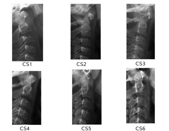
The skeletal maturity was being assessed on lateral cephalometric radiograph by using Baccetti method. 4 According to this method, the lower border of the bodies of the second (C2), third (C3), and fourth (C4) cervical vertebrae traced from the lateral cephalometric radiographs were visually analyzed and rated on a 6-level scale from cervical stage CS 1 to CS6. The patient is classified into one of six stages. (Figure 2) Pearson's coefficient was applied to measure the association between chronological and dental ages and Spearman rank correlation coefficient for the CS and dental calcification stages. Kappa test was calculated to evaluate intra-and inter-observer reading, one month after the initial assessment, 20 panoramic radiographs and lateral skull cephalograms was be reassessed in random selection, once by the same investigator and other by second investigator. Good agreement was seen between the two measurement, for dental age 1.000 and 0.89, whereas, for CVM stages, 0.864 and 0.859 respectively (intra-and inter-examiner agreement respectively).
3. III. Results
The total numbers of the studied radiograph were 112 (47 males and 65 females). It was clear that the mean chronologic age of female was significantly lower than that of male on first four stages. In stage CS3, the mean chronologic age was 12.0533±0. 976 years for females and 13.34±1.66 years for males. (Table 1) Table 2 shows the results of Spearman rank order correlation coefficients between chronological and dental ages in each cervical vertebral maturation stage. These correlation coefficients were between 0.922 and 0.177 for gender. The significance level for all coefficients was high except in CS4 in male and CS 5 in female was P> 0.5. Table 4 shows the Pearson correlation coefficient, the results revealed relationships between the chronological and dental ages; an overall high statically significant in genders was noted. Spearman rank-order correlation coefficients between the cervical vertebral maturation stages and developmental stages for the 7 teeth are given in Table 5. All correlations were statistically significant. The level of the correlation was different for individual teeth where the correlation coefficients ranged from 0.276 to 0.772 for females and from 0.453 to 0.774 for males. (Table 5) There was wide variation in tooth calcification stages for all teeth in boys and girls. The percentage distribution of dental development stages was calculated for the canines, first and second premolars, as well as the second molars. In CS1, the most frequently observed dental development stages were G for the canine and first premolar and E for the second molar (50%) in females and F for the canine and D in males (46.1%). Tooth calcification stages in other teeth had a percentage distribution less than 50%. (Figure 4) In CS2 a wide distribution of tooth calcification stages can be clearly seen stage F in the first and second premolar (80%) in female and stage F in the second premolar (60%) in male. (Figure 5) In CS4 stage G of the second (78.9%) in female and the calcification of the canine was complete in male. (Figure 7).
4. IV. Discussion
Skeletal and dental maturity assessment is a common clinical practice in many health professions especially for growth modification.
In the present study, no males were documented in stage 5 of cervical maturity stages and only two in stage 6; subsequently the mean of the chronological age in females was significantly younger than males in the first four stages, which indicated that female maturation ahead of males, which in agreement with previous studies among other population.
In current study the mean chronologic age in stage CS3 was 12.0533 + 0. 976 years for females and 13.34 ± 1.66 years for males, similar results had been recorded among both gender in Turkish and Pakistani populations 19,20 and South Africa males, 21 Indian females. 22 In contrast to the results obtained by Irfan et al, 23 Chen et al 24 and Al-Hadlaq et al 25 in Kashmiri, Chinese and Saudi populations.
High significant correlations were showed between chronological, dental ages and skeletal maturity, the highest correlation between chronological age and dental age was observed in CS 2 and CS 1 in male and female respectively. It was confirm that the cervical vertebrae stages were progressed with increase the chronological age as well as dental age.
Engestrom et al, 26 Sukhia et al 20 and Ingrid et al, 27 agreed with the present results, although Engestrom et al in Sewden samples depend on the third molar development and Ingrid et al in Central Poland samples showed the highest correlation in CS1 for both gender.
High statistic significant correlation (p= 0.000) was observed in the current study between chronological and skeletal maturity. Which mean the chronological age can be used for measuring the skeletal maturity; although not all age group were included in this study there for we cannot depend on this result unless further study prove this fact. Baidas et al 28 in Saudi Arabia and Al khal et al 25 in Southern China, 29 concluded the same results.
In the current study, the dental maturity assessment stages of Demirjian et al were used due to calcification stages of teeth as an alternative of eruption were chosen because tooth development was proposed as a more reliable criterion for determining dental maturation. A high correlation coefficient was observed between chronological and dental ages (r = 0.925, p = 0.0001for male and r= 0.845, p=0.0001 for female) in this study, which in accordance with previous results observed by Parabhakar et al , 30 Ingrid et al and Hedege et al 31 whereas a statistically significant differences between dental and chronological age were described in Belgian children due to overestimation of the chronological age with dental age, 32 and Kuwaiti children due to a tendency for delayed dental maturation. 33 The ethnic background, racial and environment influences as well as the methods, the results among populations were diverged.
A high significant correlation was observed between the dental developmental stages and cervical vertebral maturation stages of subjects with the Spearman rank order correlation coefficients.
In the present study, the permanent central and lateral incisors as well as the first molars were excluded from analysis owing to the medium correlations with CVM, whereas the canines, first and second premolars, and second molar showed good correlation, which agreed with numerous previous studies in literature, accordingly, the percentage distribution of dental development stages was calculated for the canines, first and second premolars, as well as the second molars.
In this study tooth development stages relative to stages of skeletal maturation was considered separately for male and female subjects. The sequence of each tooth according to dental development stages from the highest to the lowest correlation, the mandibular first premolar had the highest correlation coefficient with CVM stage among male subjects (r = 0.77, p = 0.001), followed by canine (r = 0.759, p = 0.001), second molar (r = 0.758, p = 0.001) and second premolar (r = 0.752, p = 0.001). Whereas for female, the mandibular second premolar had the highest correlation with CVM stage (r = 0.772, p = 0.001) followed by second molar (r = 0.756, p = 0.001), first premolar (r = 0.672, p = 0.001) and canine (r = 0.586, 0.001). This finding was confirmed that the first premolar and second premolar recorded with highest correlation in male and female respectively. Similarly study among Chinese, the second molar and canine for female and male respectively, 24 while in Saudi male revealed higher correlation values in the first premolar and the second molar with the skeletal maturation. 25 Moreover Krailassiri et al obtained the same finding among female, while the highest correlation in the second premolar among male. 34 In this study stage F in the mandibular first premolar was 38.4% in CS1 and 37.5% in CS2, and in stage H was 62.6% in CS3, 88.9% in CS4 and 100% in CS6 in male. Whereas in female the percentages for stage F in mandibular second premolar were 25% in CS1, 80.0% in CS2 and for stage G 73.4% in CS3. For stage H, the percentages were 52.6% in CS4, 93.7% in CS5 and 100% in CS6.
In study carried out by Uysal et al, among Turkish population showed that the development of canine and first premolar was completed in most cases at the pubertal growth spurt. 19 However in Chinese and Indian populations, stage F in second molar was observed in female at beginning of the pubertal growth spurt. 22,24 This differences may be partially related to discrepancies in the, age, racial background and
5. V. Conclusion
? A statistically significant relation was observed between chronological and dental ages and dental developmental stages with skeletal maturity stages. ? The stage H of the first premolar in male and stage G of the mandibular second premolar in female suggest the beginning of the pubertal growth spurt in Sudanese subjects. This confirms that the first premolar and second premolar (male and female respectively) may be used as markers for skeletal maturity of a child who's seeking orthodontic treatment.
Volume XVI Issue I Version I


| Cervical vertebra | Male | Female | Total | ||
| stages CS | No | Mean , SD | No | Mean, SD | |
| CS1 | 13 | 9.84+1.55 | 4 | 9.42+2.02 | 17 |
| CS2 | 8 | 11.83+2.13 | 5 | 9.7+0.9 | 13 |
| CS3 | 16 | 13.34+0.97 | 15 | 12.05+1.66 | 31 |
| CS4 | 8 | 14.73+0.76 | 19 | 13.29+1.01 | 27 |
| CS5 | 0 | 0 | 16 | 15.03+0.65 | 16 |
| CS6 | 2 | 15.45+0.55 | 6 | 15.35+1.9 | 8 |
| Total | 47 | 65 | 112 |
| CS | Male | Female | ||
| R | P | r | P | |
| CS1 | 0.864 | 0.000 | 0.922 | 0.000 |
| CS2 | 0.915 | 0.001 | 0.759 | 0.010 |
| CS3 | 0.764 | 0.001 | 0.552 | 0.030 |
| CS4 | 0.177 | 0.675 | 0.731 | 0.000 |
| CS5 | 0 | 0 | 0.239 | 0.373 |
| CS6 | 0 | 0 | 0.912 | 0.050 |
| P value was significant at < 0.05 | ||||
| Gender | R | P |
| Male | 0.744 | 0.000 |
| Female | 0.878 | 0.000 |
| P value was significant at < |
| 0.05 |
| Tooth | Male | Female | ||
| R | P | r | P | |
| Central incisor | 0.453 | 0.001 | 0.276 | 0.026 |
| Lateral incisor | 0.408 | 0.001 | 0.451 | 0.001 |
| Canine | 0.759 | 0.001 | 0.586 | 0.001 |
| 1 st premolar | 0.774 | 0.001 | 0.672 | 0.001 |
| 2 nd premolar | 0.752 | 0.001 | 0.772 | 0.001 |
| 1 st molar | 0.496 | 0.001 | 0.425 | 0.001 |
| 2 nd molar | 0.758 | 0.000 | 0.756 | 0.001 |
| Total | 0.704 | 0.001 | 0.769 | 0.001 |