1. I. Introduction
ung cancer is the leading cause of cancer-related mortality around the world, with 85% of cases identified as non-small-cell lung cancer (NSCLC) 1. The epidermal growth factor receptor (EGFR) signaling pathway plays a crucial role in regulating tumor growth and survival and is important in the progression of NSCLC 2 . The signaling pathway of EGFR is estimating to be activated in more than half of patients with NSCLC, increasing the role of targeted therapy. 3 Two reversible EGFR tyrosine kinase inhibitors (EGFR-TKIs), gefitinib and erlotinib, are often used therapies in EGFR-mutated NSCLC and are approved by the US Food and Drug Administration 4,5 . They are also approved for treatment of carcinomas of the pancreas that are locally advanced. Treatment is often well tolerated, with mild common adverse side effects of skin rash and diarrhea. 6 However, growing incidence of major adverse side effects, such as interstitial lung disease (ILD), continues to be reported since drug approval 7,8 . Though the mechanism of adverse-pulmonary events is not completely understood, careful recognition and documentation of both incidence and risk of ILD is important to allow early recognition of potential toxicity.We report a histologically confirmed case of fatal interstitial pneumonitis, with acute lung injury, associated with erlotinib therapy.
2. II. Case Report
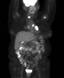
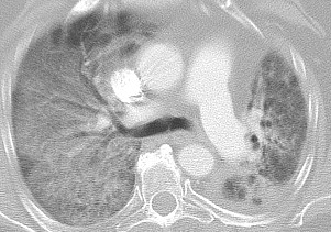
A 68-year old female, non-smoker, with obstructive sleep apnea requiring bi-level positive airway pressure, chronic obstructive lung disease, and noninsulin dependent diabetes mellitus was diagnosed with metastatic adenocarcinoma of the lung (to left humerus and thyroid). She initially presented with a 10-year history of asymptomatic hypercalcemia for which imaging of the parathyroid gland was performed and incidentally showed several abnormal lung masses. Subsequent staging scans showed a large hilar mass as well as a large mass in the left lower lobe, several metastatic lesions in the left lung, and a lytic lesion in the left humerus (Figure 1,2). She underwent VATS with wedge resection of the lung nodules for diagnostic purposes. Pathology showed poorly differentiated adenocarcinoma and genomic testing revealed a mutation in EGFR. She completed a short course of palliative radiation therapy to the primary tumor in the lung to prevent bronchial compression as well as to the humerus for pain control. She initiated systemic therapy with erlotinib 150mg PO daily, which she started midway through her RT course.
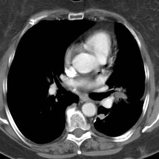
Two weeks following the beginning Erlotinib treatment (4 weeks following radiation therapy), she reported a dry cough and dyspnea on exertion. Her pulse ox dropped to 92% after ambulation. A CT scan showed smaller masses and scarring in the left lower lobe in the area of surgery (Figure 3).
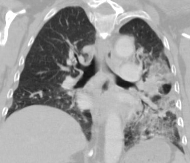
Approximately one month later, she reported increased SOB without hemoptysis, fevers, or chills. She was treated with a prednisone taper, initial dose of 60mg daily. The patient transiently responded well. However, upon lowering the daily dose to 40 mg her symptoms worsened, and a 60 mg dose was resumed. Repeat CT scan showed smaller masses, but new left lung infiltrates and an area of consolidation in the left lung that vaguely outlined the radiation port. There was an associated left pleural effusion (Figure 4). Follow-up visit showed resting oxygen level of 88%. She was prescribed home oxygen therapy and levofloxacin for bilateral pneumonia. Despite the initiation of antibiotics and steroids, the patient reported worsening of her symptoms, with SOB at rest, persistent cough, and new onset fevers and chills. She was admitted to the hospital.
On admission, vital signs were significant for a temperature of 100.4 F, heart rate of 116, respiratory rate of 16, and oxygen saturation of 70%. Blood gas showed pH 7.48, pCO2 28 mmHg, pO2 <44mgHg, O2 saturation 75.4%. Labs included hemoglobin 11.7 g/dl, WBC 17.5 thousand/uL with left shift. Her creatinine was at baseline. CTA of the chest was negative for pulmonary embolus, but noted significantly worsened airspace opacities bilaterally when compared with prior films. Patient was placed on broad-spectrum antibiotics and transitioned to prednisone 40 mg twice daily. Blood cultures revealed no organisms. Follow-up chest x-ray demonstrated diffuse bilateral infiltrates and pulmonary edema. Echocardiogram was unremarkable for a cardiac etiology of her symptoms. On day three of admission, patient required intubation for respiratory distress and vasopressor therapy for hypotension.
A bronchoscopy was performed. Bronchial washing was negative for diffuse alveolar hemorrhage. New labs revealed hyponatremia. Respiratory viral panel, Cytomegalovirus, Pneumocystis, Legionella and other infectious testing were negative. Her respiratory distress increased and steroids were thus increased to 60mg every six hours. Despite maximal medical and ventilatory support, the patient continued to deteriorate rapidly with increased oxygen requirements and worsening hypoxemia. Repeat CT chest revealed bilateral, diffuse pneumomediastinum with posterior displacement of the heart, and worsening airspaces, sparing the apices (Figure 5). Patient's condition deteriorated so she was transitioned to comfort care and expired.
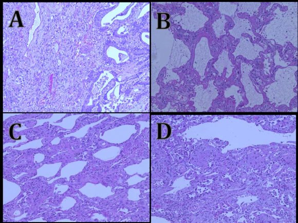
A limited autopsy of the lungs was performed. Gross examination revealed pale lungs with cobblestoning. Microscopic examination (Figure 6) showed varying stages of diffuse alveolar damage (DAD).The right lung exhibited diffuse, early to acute organizing proliferative DAD. The left lung exhibited acute organized proliferative DAD, with end-stage DAD over the left lower lobe. There was no evidence of residual tumor. These findings led to a clinical diagnosis of interstitial pneumonitis due to erlotinib treatment, likely superimposed with radiation pneumonitis. The different stages of DAD within various lobes of the lung, with severe honeycombing over the irradiated lobe suggest interstitial pneumonitis as a complication of erlotinib treatment in the setting of overlying radiation pneumonitis.
3. III. Discussion
New cancer therapies are quickly emerging, with promising, effective treatments for many patients.
Erlotinib received FDA approval in 2004 as monotherapy for locally advanced or metastatic NSCLC after failure of at least one prior chemotherapy regimen. In the original FDA Drug Approval summary, ILD was observed during treatment in 0.8% of patients; similar to the placebo incidence. 3 The EGFR-TKIs are thought to be relatively safe therapies in the treatment of patients with advanced NSCLC. Although reported with a relatively low incidence ranging from 3.5-5%, ILD is a growing concern as a significant, sometimes fatal adverse side effect. 9 Assessment of ILD is particularly challenging, as it is not a single disease entity, but a spectrum of lung pathology. In addition, patients with NSCLC often have confounding factors, such as heart failure, pulmonary infectious disease, and/or disease progression within the lungs. In addition, the pathophysiology of drug-induced injury is not well understood. Some have suggested that EGFR inhibitors interfere with the repair process of pulmonary tissue, as epidermal growth factor is secreted onto bronchial surface fluid by pneumocytes. The primary damage is thought to occur due by immunocyte activation, likely modulated by a number of host and environmental factors.
It is particularly important to highlight the risk and incidence of ILD as EGFR-TKIs are increasingly used in daily practice, both for their efficacy and tolerability. Identifying patients at high risk of ILD important to reduce occurrence. Unfortunately, there are currently no guidelines for treatment as randomized controlled studies investigating the issue are lacking. For now, providers are suggested to discontinue treatment in patients with known ILD. 3-4 10 Drug associated ILD appears to be higher among Japanese patients than Caucasian patients for reasons unclear. 4 No study has yet examined the relationship between constitutional or environmental factors specific to Japanese Patients. ILD related to drug therapy requires high suspicion and exclusion of other potential causes.
Certainly, more studies are needed to understand the disease process.
Common histopathologic studies reveal alveolar edema, pneumocyte hyperplasia, the accumulation of fibrin, and formation of hyaline membranes. Of note, these findings are not pathognomonic, as they are also seen in acute-respiratory distress syndrome and acute interstitial pneumonitis. 11 The time range between drug administration and symptom presentation is highly variable, ranging from 2-282 days. 12 Diffuse alveolar damage (DAD) is a progressive process that goes through several stages. The early acute stage exhibits edema and hyaline membrane formation over the alveolar cells. Subtle interstitial fibrosis may be seen. The early stage develops within the first 1-2 weeks. With progression, the organizing fibroproliferative stage develops. Hyaline membranes are being actively resorbed, and fibroblasts migrate into the interstitium. Fibrin thrombi are seen within the vessels. There is acute inflammation, squamous metaplasia, and type 2 pneumocytes proliferate to replace the damaged alveolar cells. 13 As the disease process progresses, end stage DAD ensues, characterized by increased squamous metaplasia, marked fibroblastic proliferation, bronchiolar dilatation, and severe fibrosis of the interstitium. The end result is a solid honeycombed appearance of the lung. Clinically, patients present with dyspnea and cough, and as the disease progresses, work of breathing is increased and patients eventually develop respiratory failure, as seen in the case presented. Radiographically, there are non-specific changes, manifested by edema and diffuse, infiltrative opacities of the lung. Treatment is with corticosteroids and supportive care. Definitive treatment is with lung transplant. 14,15 Diffuse alveolar damage may be instigated by different factors, and a thorough history is required to find causal source. Acute radiation pneumonitis affects 10% of patients who undergo radiation therapy. Clinical manifestation is usually evident 1-6 months after radiation treatment. X-rays show diffuse chest infiltrates over the area of the radiation portal. While this may spread to non-irradiated areas of the lung, bilateral involvement is rare. 16 Erlotinib and radiation may have a synergistic effect, increasing the incidence of interstitial pneumonitis in patients treated with both. A study of patients who developed interstitial pneumonitis with concurrent treatment with radiation therapy and erlotinib showed the pneumonitis could not be reversed with methylprednisone. 17 Identifying at-risk patients is a major concern that warrants further investigation. At this point, more is known regarding potential risk factors for gefitinib. A retrospective survey of the prevalence and risk factors for gefitinib-induced ILD in Japan found ILD to be significantly associated with male gender, prior smoking history, and concomitant interstitial pneumonia. Prior chemotherapy and radiation to the lungswas also reported as a predisposition to ILD. 18 Patients on erlotinib often have already received treatment with other antineoplastic agents. Pulmonary toxicity, ranging from interstitial pneumonitis to acute respiratory syndrome, is not an uncommon side effect of chemotherapy, as it has been described for gemcitabine Whether these findings are also correlated with erlotinib is yet to be studied.
4. Volume XVI Issue V Version I
5. 19
, mitomycin 20 , vinorelbine tartrate 21 , docetaxel 22 , ifosfamide 23 To date, pre-existing pulmonary disease is not an absolute contraindication to treatment with erolitinib.
Given the growing number of reported pulmonary toxicities, the authors recommend documenting baseline respiratory status and symptoms prior to the initiation of medication. This would allow objective monitoring of subtle changes after initiation of drug therapy.
. Lung injury occurs usually early after administration of chemotherapy. Whether erlotinib itself contributes to lung toxicity due to its own unique chemical properties or rather exacerbatespreexisting pulmonary toxicity from prior chemotherapy or radiation is yet clarified.
Limitations at this time are extended to management of erlotinib associated lung toxicity. Case reports show varied success with high dose corticosteroid therapy. Though many patients seem to have responded to supportive therapy (including high dose supplemental oxygen and mechanical ventilation when necessary), many patients have died due to progressive respiratory failure, as seen with the patient whose story is described above. In addition, follow-up is limited when evaluating for recurrence of pulmonary disease in those with him authors have described success.
In conclusion, as demonstrated, EGFR-TKIs carry the substantial risk of developing ILD in patients with advanced NSCLC, although infrequently. Awareness of the potential for lung toxicity is necessary. Erlotinib should be considered among the antineoplastic agents with the potential to contribute to pulmonary disease. Physicians are encouraged to evaluate new or worsening pulmonary symptoms in patients receiving EGFR-TKI therapy. Further studies are needed to better elucidate risk factors, disease pathophysiology, and potential treatments to lower the incidence and mortality of ILD associated with EGFR-TKIs.