1. I. Introduction
horiocarcinoma is extremely rare and aggressive form of gestational trophoblastic disease. It occurs due to neoplastic changes in the chorionic villi epithelium. The most common site of origin is the uterus; the incidence of choriocarcinoma in the fallopian tube is very low [1]. We report a case of choriocarcinoma of the Fallopian tube treated successfully with surgery and chemotherapy.
2. II. Case Report
The patient is a 37 years-old woman, admitted to emergency room vaginal bleeding and lower abdominal pain without amenorrhea. Her menstrual cycles was regular; she had 2 vaginal deliveries and no previous gynecologic operations.
On admission, her temperature was 37°C pulse rate 90pulse/min, and blood pressure 125/70 mm Hg. Vaginal examination revealed moderate vaginal bleeding and a normal ante flexed uterus, with no adnexal palpable tumor. Her blood test results were: Hemoglobin 12, 7 g/l00 ml of; platelets 500.000/mm3; her blood group A+. The patient underwent transvaginal ultrasonography which showed a normal uterine cavity with no signs of intrauterine gestational sac or embryo. But an ectopic mass of 49x36 mm was visualized in the right adnexal region, vascularized with Doppler flow, and there was images of free fluid limited to the pelvis. A level of 15943 mIU/ml ?-Hcg was detected.
Author ? ? ? ?: Service Gynéco-Obstétrique, cancérologie et Grossesse à haut risque Maternité Souissi. RABAT. e-mail: [email protected] An emergency laparotomy was made. On exploration, was noted an abundant haemoperitoneum with 700 cc of blood that was evacuated, on the right fallopian tube as well was an actively bleeding ectopic mass invading the right lateral side of the uterus; ovaries weren't affected, interadnexial subtotal hysterectomy was performed. The postoperative course was uneventful.
Histological examination of the specimen revealed an invasion of the fallopian tube with signs of rupture. No villous formations were found, the uterus was partially invaded. A diagnosis of choriocarcinoma of Fallopian tube was made. The patient underwent complement of radiologic and laboratory examinations. Computed tomography scans of the abdomen and thorax were realized showing Pulmonarmetastasis. The patient workup indicated that the FIGO score was 6. Chemotherapy was planned, she was administered monochemotherapy using methotrexate 1 mg/kg intravenously on days 1, 3, 5 and 7 alternating with folinic acid 0.1 mg/kg intramuscularly on days 2, 4, 6 and 8 of the cycle.
Treatment response was monitored weekly by checking serum ?-Hcg measurements that gradually decreased and became negative after 2 months and then monthly measurements were made. The patient responded well to the chemotherapy and no side effects were observed. The patient was followed up for 1 year and was disease free till that period.
3. III. Discussion
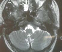
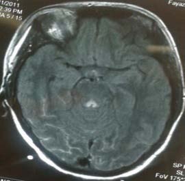
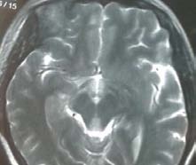
Choriocarcinoma is a malignant type of the gestational trophoblastic disease which usually originates from uterine cavity. On rare occasions they may occur in the tube, cervix [2] horn of the uterus, vagina or other pelvic organs [3]. The incidence is 0.8% of all the gestational trophoblastic disease [4]. It is generally very aggressive [5]; and otherwise can simulate other gynecologic diseases such as ovarian cyst, tubo-ovarian abscess and ectopic pregnancy [4], with clinical similar symptoms as amenorrhea, vaginal bleeding and vascular instability along with increased B hCG titer. MRI, endovaginal ultrasound and colourflow Doppler play an important role in the diagnosis of intrauterine choriocarcinoma [6]. However, no specific Year 2017 imaging findings have been defined for extra-uterine choriocarcinoma; in our case, the ultrasonography suspected malignant adnexal tumor but it wasn't visible with MRI.
Histological examination of a surgically resected specimen is essential for the confirmation and diagnosis of choriocarcinoma. In our case, a tubal pregnancy was suspected, but the examination of the specimen affirmed the diagnosis of choriocarcinoma.
Choriocarcinoma, produces up to hundred times the amount of ?-Hcg .?-Hcg level measured in our case was up to15943 mIU/ml ?-Hcg. Therefore, the measurement of ?-Hcg concentrations are very useful to assess the response to treatment and detect any recurrences.
The treatment of tubal choriocarcinoma is as of the uterine type, surgery with chemotherapy [7]. In tubal choriocarcinoma conservative treatment for younger women can be established. Salpingectomy or adnexectomy without removal of the uterus Is done, followed by chemotherapy [8]. For older women with no child desire, a bilateral adnexectomy or hysterectomy can be preconized [9]. Our Patient underwent hysterectomy.
Choriocarcinoma can metastasize into the lungs, brain, liver, and even very rarely into the fetus [5].
Because of this highly metastatic potential , monochemotherapy using méthotrexate and l'actinomycine D ; or multiple drugs chemotherapy using méthotrexate, d'actinomycine D, d'étoposide, cisplatine, cyclophosphamide, vincristine and bléomycine [8] is essential and proves to be very effective in trophoblastic tumors. The literature shows that patient with choriocarcinoma even with metastasis can achieve complete remission [10].
The women who are treated for extra-uterine choriocarcinoma should receive effective contraception for 1 to 2 years after the completion of their treatment, with B-hcgmonitoring.
4. IV. Conclusion
The tubal choriocarcinoma is a rare disease with bad prognosis if not treated. This study reminds us the importance of the histological examination of any ectopic pregnancy. The tubal choriocarcinoma diagnosis can be made while suspecting an ectopic pregnancy with a high level of ?HCG, a salpingectomy must be carried, and chemotherapy must be followed to improve the prognostic and surviving rate.