1. I. Introduction
astroschisis is a congenital defect in the anterior abdominal wall right to the umbilical cord resulting from incomplete closure of the lateral folds during sixth weeks of gestation 1 . As a result the small bowel and other viscera are exposed to amniotic fluid until delivery and to environment after. Incidence varies from 1 in 4000 to 1 in 10000 live birth 2 and this is increasing world wide 8 . In contrast to omphalocele associated anomalies are infrequent 3 . The outcome of neonates with gastroschisis has improved over past decades. Though most series claims survival rates over 90% 4,5,6 , our experience is still frustrating. Several factors are associated with adverse outcome in gastroschisis, including prematurity, low birth weight, absence of prenatal diagnosis, place of delivery, timing of repair, type of repair, associated anomaly and sepsis 5,6 . The aim of this study was to evaluate the outcome and identify the factors influencing the outcome and how to address these factors to improve outcome in gastroschisis in a tertiary care pediatric surgery center of Bangladesh.
2. II. Materials and Methods
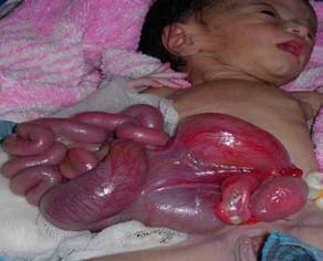
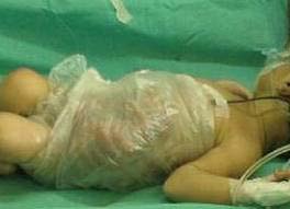
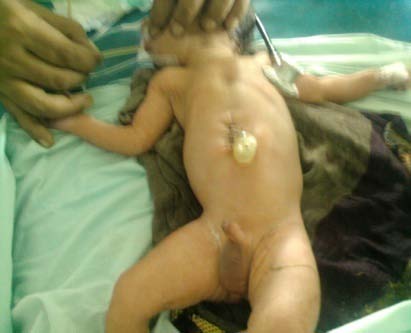
It was a retrospective analytical study done in Dhaka Shishu (Children) Hospital from March 2014 to April 2017. Hospital records of all patients with gastroschisis were reviewed. Immediately after admission, exposed viscera (photograph 1) were covered with plastic bag (photograph 2). Patients were covered with cotton sheet, kept nothing per oral; an 8Fr feeding tube was inserted for nasogastric suction. Intravenous fluid resuscitation and antibiotic started immediately. All patients received injection vitamin K. investigations performed on admission were blood grouping and Rh typing, random plasma sugar, serum electrolytes. After initial resuscitation patients were taken to Operation Theater and reposition & primary repair tried under general anaesthesia (photograph 3). When reposition was not possible, silo was performed with sterile saline bag or urobag (photograph 4). After operation patients kept nothing per oral, NG suction and intravenous nutrition maintained until abdominal distention reduced & bowel movement established. Silo was squeezed every alternate day and repair performed when complete reposition was possible. Neonates were divided into two groups. Group A (Neonates who survived), Group B (Neonates who expired). Data were collected regarding prenatal diagnosis, gestational age, birth weight, place of delivery, associated anomaly, time from delivery to surgery and final outcome. Ethical clearance was taken from hospital ethical committee. Statistical analysis was done using SPSS Version 22 software. Associations of continuous data were assessed using student t-test. Associations of categorical data were assessed using Chi-square test and Fisher's exact test. For both test, p<0.05 was considered significant.
3. III. Results
Out of 75 neonates admitted during the study period, 43 were male and 32 were female (figure 5). Only 14 patients survived (18.7%). Prenatal diagnosis was done in only 3 patients among whom 2 patients survived. Mean gestational age was 35.71±1.06 weeks in group A and 34.34±1.42 weeks in group B. This difference was statistically significant. Mean birth weight was 2.19±0.14 kg in group A and 2.00±0.20 kg in group B. Eight patients out of 14in group A were delivered within Dhaka division in group A and only18 patients out of 61in group B were delivered within Dhaka division. Six patients had associated intestinal atresia, all of them expired. Mean time from delivery to surgery in group A was 13.14±2.41 hours and in group B was 18.75±3.86 hours. Silo performed in 40 patients. Among them only one survived. Thirty five patients had primary repair of which 13 survived (table 1).
4. IV. Discussion
Pediatric surgery division of Dhaka Shishu (Children) Hospital is the largest tertiary care pediatric surgery center in Bangladesh. With limited resource we are continuously trying to improve our service. When it comes to gastroschisis, we are still struggling. So, we tried to find out where to focus.
We found more male patients than female in this study. Owen A et al. found same 7 but Bradnock T J et al found opposite 8 . According to Klein M D gastroschisis occur predominantly in male but definite explanation yet to found 3 .
Prenatal diagnosis is believed to improve outcome in gastroschisis by optimizing time, place and mode of delivery. In most reported studies prenatal diagnosis significantly affected outcome 2,6,8 . But we found only 4% (3/75) patient with prenatal diagnosis. This is due to less public awareness about prenatal care & screening in a developing country like Bangladesh. Limited experience of radiologists and primary care giver at rural area might also be a contributing factor. In advanced centers prenatally diagnosed cases are delivered in regional centers and after delivery, herniated bowel immediately placed in plastic bag to prevent hypothermia and hypovolemia 6 . Quirk J G et al demanded resuscitation and stabilization of neonates with gastroschisis by an experienced team of neonatologists. Advantages include the prevention of hypothermia, hypovolemia and assurance of nasogastric drainage 2 .
Quirk J G et al showed delivery in the regional center is associated with the better outcome 2 . We found same result. This is due to early transport and closure of the defect. Fasching G et al & Quirk J G et al reported no significant difference in outcome with mode of delivery 2,11 . Time from delivery to surgery is crucial. In this study we found huge difference with most reported studies. Most authors urges earliest possible repair of gastroschisis and they do it within 5 hours of delivery 2 . In our center it is much delayed as most of the babies come from outside Dhaka. When we received the neonates the exposed viscera were already swollen and edematous and patients were in severe hypovolemia & hypothermia. It further delays the surgery and made reposition very difficult. Hence silo was performed in most patients though primary repair is treatment of choice 5,6 . Several studies reported better outcome using preformed silo 4,7 . We used sterile saline bag or urobag to form a silo. Almost all of this patients developed sepsis & were associated with poor outcome.
Mean gestational age & birth weight was significantly higher in survivor group. This finding is similar to most of the series 4,5,6,8 .Fasching G et al however showed gestational age has no influence on outcome. This is because of advanced neonatal intensive care (NICU) and nutritional support 9 . In our center NICU support is not always available for these babies and it is very difficult to manage these premature low birth weight babies in ward. Most of them suffer from hypothermia, sepsis and acidosis. Watanabe et al & Calcagnotto et al reported low birth weight in gastroschisis as a factor for increased mortality 9,10 .
Intestinal atresia is another poor prognostic factor in neonates with gastroschisis. In this series we found 83.33% mortality (5/6). Driver et al & Snyder et al reported increased morbidity but not mortality in these cases 5,6 . This is may be due to advanced NICU support & facilities for prolonged parenteral nutrition, which are not available in our setup.
5. V. Conclusion
Factors adversely influence the outcome are absence of prenatal diagnosis & planning of delivery, prematurity & low birth weight, associated intestinal atresia and duration from delivery to surgery. Immediate resuscitation & covering of exposed viscera after delivery is also of great importance. Efforts must continue to raise awareness among general people as well as among obstetricians to increase prenatal diagnosis and to instruct newborn care providers in peripheral hospitals in the appropriate initial care of these high risk neonates. Neonatal surgical intensive care is a crucial factor as almost all neonates are premature.
6. Global
| Group A: Survived | Group B: Expired | ||
| (n=14) | (n =61) | ||
| Prenatal diagnosis | 2 | 1 | |
| Mean gestational age (weeks) | 35.71±1.06 | 34.34±1.42 | .001 |
| Mean birth weight (kg) | 2.19±0.14 | 2.00±0.20 | .001 |
| Place of delivery | within Dhaka= 8 | Within Dhaka=18 | |
| Outside Dhaka=6 | Outside Dhaka= 43 | ||
| Mean time from delivery to | 13.14±2.41 | 18.75±3.86 | .001 |
| surgery (hour) | |||
| Intestinal atresia | 1 | 5 | |
| Type of surgery | Repair=13, Silo=1 | Repair=22, Silo=39 | .001 |