1.
hird most common malignant tumor in the salivary glands is Adenocarcinoma which accounts for less than 1% of all malignancies and for from 5% to 20% of all carcinomas in the sinonasal area. 1 Few reports in the literature describes the diagnostic imaging findings of adenocarcinoma arising in the maxillary sinus. They are painful, fast-growing masses, but occasionally present as painless and slow-growing.
2. II. Case Report
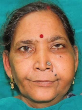
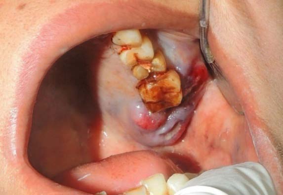
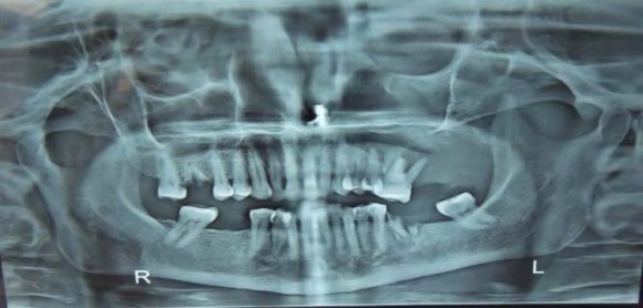
A 48-year-old woman was referred to our hospital with the chief complain of swelling in the left upper facial region since2-3 months. Patient did not gave any positive history of any deleterious habit. On extra oral examination a soft, elastic mass of 29mm in diameter extending anteroposteriorly from the lower left eyelid till the inferior border of mandible and superoinferiorly extending from the left corner of mouth till the tragus of ear was seen. The overlying skin was normal as surrounding skin with a slight raise in temperature (Fig 1). On palpation the swelling was soft in consistency was compressible and tender. Patient also complained of the nasal congestion and left sub occular tension. While palpating neck region a single lymph node of approx. 12mm in diameter was palpated at left level II region. On intra oral examination an ulcerative lesion was seen in the left maxillary arch region in relation to 27 28 with bluish discoloration around the ulcerative lesion. On checking the mobility status of 26 it was grade 3 mobile ( The mass compressed the orbit base, but the orbit's contents were not affected. The maxillary tuberosity and the hard palate were destroyed. The CECT images also suggested that it was not a Squamous cell carcinoma but a malignant tumor.
After the radiological the biopsy was performed which revealed parakeratinized stratified squamous epithelium overlying fibrocellular connective tissue stroma. The epithelium showed dysplastic features such as increased N: C ratio, prominent intercellular junctions, prominent and increased number of nucleoli, mitotic figures. Focal areas of invasion in the form of discrete tumor islands are seen. The C.T. stroma shows solid tumor mass areas with variable organisations formed by large polygonal cells having pale to eosinophilic cytoplasm and nucleus ranging from hyperchromatic to vesicular. Tumor cells were also arranged in forms of sheets, clusters, pseudoductal patterns with secretory material. Few cells also showed individual cellkeratinization, with numerous atypical mitotic figures. Tumor necrosis and perineural invasion was also observed and a diagnosis of Adenocarcinoma NOS (High grade) was given.
After the conformational biopsy report, surgical resection was done.
In the treatment tumors which have broad invasion into the maxillary sinus the Dieffenbach-Weber-Fergusson incision modified by Zange is used to perform a hemimaxillectomy (Fig 6).Before beginning the process of incision the area was marked and infiltrated with1% xylocaine with 1 in 100,000 units adrenaline.After the incision cheek flap was elevated from the antero lateral surface of maxilla in the subperiosteal plane. Then dissection was slightly altered so that the involved skin overlying the anterolateral wall of maxilla was also removed ebloc along with the tumor. The lymph nodes upto level V were removed. Infra orbital floor defect was reconstructed by rotating temporalis muscle flap and facia lata graft was harvested (Fig 7).
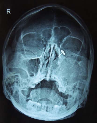
The PNS view of the skull showed radiopacity in the left maxillary sinus, nasal cavity. The radiopacity was also appreciated in the left orbit with absence of a left maxillary tuberosity line and the lower border of left orbit. At this point, a malignant tumor was strongly suspected (Fig. 4).
After the basic radiographic examinations the patient was advised for the Contrast Enhanced Computed Tomography (CECT). The coronal sections of CECT shows a well-defined hypodense lesion in the left maxillary region extending into the left maxillary sinus, left nasal cavity and left orbit. The hypodense mass is also infiltrating into the buccinators and the masseter muscle involving the massetric space. Massive destruction of the alveolar bone, walls of maxillary sinus and inferior orbital wall can also be appreciated. (Fig 5). Tensor fascia lata flap is a myofasciocutaneous flap that has been first described by Wangensteen in 1934 for abdominal wall reconstruction. This flap started to gain popularity after further description by Nahai, et al., 1978 and 1979. It has a significant role in the management of pressure sores, facial reanimation. Also, it has been used as a free flap in head and Neck reconstruction. In our case fascia lata flap was used for the reconstruction of the infra orbital floor.