1. Background
etal bovine serum is the most widely used serum for animal cell culture, mainly due to its high concentration of growth factors and low concentration of immunoglobulins. The use of FBS is associated with challenges such as high variability in batch-to-batch composition, risk of transmitting bovine infections or the initiation of the xenogeneic immune response to bovine antigens and rising cost and complications of the purification of products (Brindley et al., 2012-Bieback, 2013).
Commercially available sera, adult bovine and newly born calf serum (NBCS), sheep, horse, goat sera found to be suitable for the growth of most cell lines and primary culture. Primary cultures from G .pig, chicken embryo fibroblast, monkey kidney (Savant, 1987), peritoneal macrophages were prepared and proliferate in media with goat serum (GS) (Paranjape, 2004). Also, GS used in studies different nutrients & cellular metabolism (Parap, 1995) and virus replication (Paranjape & Cadam 1985). Growth media supplemented with GS were used for in vitro cultivation of Thielaria parasite (Sharma et al., 1998). Developments towards reduced and serum-free media, chemically defined media, maintenance media, and suspension culture media for anchorage-dependent cell lines have been realized (Tezel and Priore 1998).
A new cell culture supplement, platelet lysate, was evaluated compared with fetal bovine serum (FBS), an established industrial medium for animal cell culture. Generally, platelet lysate medium was less complex than FBS. Platelets considered as concentrated supply of platelet-derived growth factor (PDGF) and transforming growth factor (TGF) (Fukimizu and Grinell 1990) which play an important role in the growth of cell culture as they are participating in many diverse cellular functions. Also, Platelets are the primary source of a number of growth factors, attachment factors (fibronectin and vitronectin), enzymes, serotonin and other factors (Ross and Raines, 1990). The platelet lysate medium demonstrated lack of microorganisms, mycoplasma, and endotoxins (Liselott et al., 2003). There was no significant difference in DNA methylation profiles of Human Platelets lysate and fetal calf serum on mesenchymal cells and did not affect their differentiation potential towards osteogenic or adipogenic lineage (Fernandez-Rebollo et al., 2017) Thus, the goal of this study, to demonstrate the potential role of newly born calf serum, platelet lysate alone and goat serum, as a growth-promoting supplement in media used for the growth of different cells line culture (Vero & MDBK).
2. II.
Materials and Methods a) Collection of goat blood (According to Paranjape, 2004) Keep muslin clothes covered bucket & gathered the blood as soon as the animal slaughtered. Poor the blood and leave to clot at room temperature then leave at 4 0 C overnight. Discard the Cotton muslin, poor the serum and centrifugate 300 rpm for 30 min at 4 0 C. Filtration by sietzfilter, sterility was applied then the serum was inactivated at 56 0 C for 30 min. Quality control testing for the presence of antibodies.
3. b) Equine growth factors E-GF TM
E-GF TM is a preparation consisting of a pale yellow round cake of Lyophilized equine Platelets Growth Factors E-GF TM According to Schallmoser and Strunk, (2013). Blood (800-900 ml) collected from 5 mature horses; blood centrifuged at 200 g for 15 min. The plasma was then centrifuged at 400 g for 15 min. The platelet pellet then resuspended with aspirated platelet-poor plasma. The PC diluted to 1 x 10 12 platelets/ l after a complete blood platelets count was performed. Platelet lysate then generated by a single freeze/thaw Cycle at 80°C overnight followed by 37°C thaw. Resulting products were pooled, centrifuged at 4000 g for 15 min, and filtered with 0.22 lm filters and was stored frozen at -18 0 C ready for use. The breakdown of platelets in vitro before lyophilization leads to release of supra-physiological doses of growth factors. The vial is reconstituted in 2 ml of sterile normal saline; gently swirl the vial for 3 minutes just before use in cell cultures. The reconstituted vial shouldn't be used after 1 hr.
4. c) Cellular toxicity
In 96 wells T. C plate in triplicates wells, dispense100µ of either Vero (African green monkey transformed kidney epithelial cells) and (MDBK) Madien Darby Bovine Kidney cells cell lines supplemented with different concentrations (4, 6 ,8, 10, 12 & 15 %) of Growth enhancing additives (GS, EPL, and NBCS). Incubation was performed at 37 0 C for 2-3 days with daily examination under a light microscope.
5. d) Cell cultures and Viability
Vero & MDBK cells were cultured in Modified Eagle's Medium (MEM) with 0.1% Penicillin-Streptomycin, in duplicates T.C-flasks for each concentration and incubated at 37 0 C. Growth promoting additives (GPA) added to media in concentrations 4
6. Results
7. a) Cellular toxicity
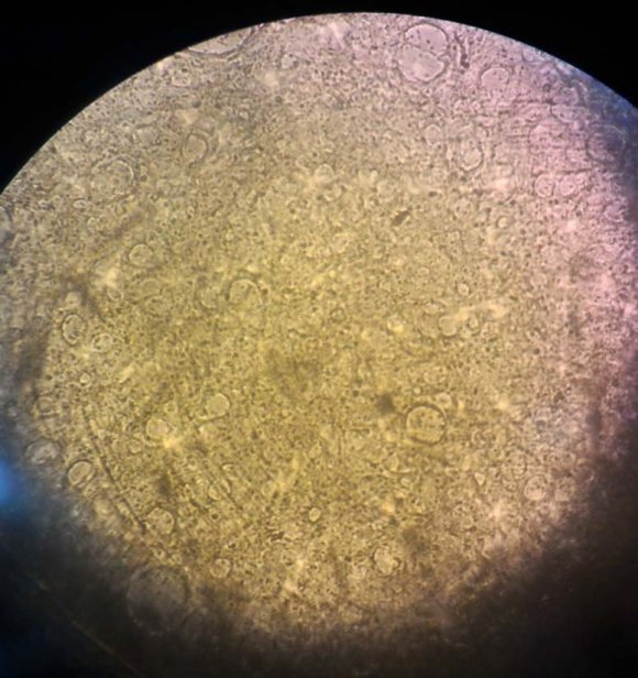
Vero & MDBK cell lines cultured in medium supplemented with GS (Table , 1) showed No toxicity at all concentrations (6, 8, 10, 12 & 15 %) with the preferable growth rate. Low Concentration of Growth factors at 4% showed abnormal cellular shapes, but these changes did not end with cell death so all tested concentrations can use safely.
The previous cell lines exhibit No toxicity at all concentrations (4, 6, 8, 10, 12 & 15 %) using platelet lysate.
8. b) Proliferation and viability assays
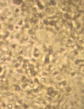
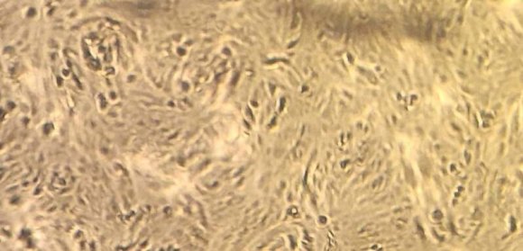
Goat serum exhibit flattened and irregular heterologous shape of cells in the first passage, after that adaption of cell lines occurred in the following passages. The results of figure (1) revealed that cells had been proliferated using goat serum in concentration 5% & 6% in MDBK and Vero cells. 0.5-1% Equine Platelet lysate (EPL) could be added to growth mediain case of low concentration of (GS) that induced a preferable cellular morphology and proliferation rate. Growth rate% in cell lines recorded 80 and 90% in Vero & MDBK cells respectively at 7% of goat serum.
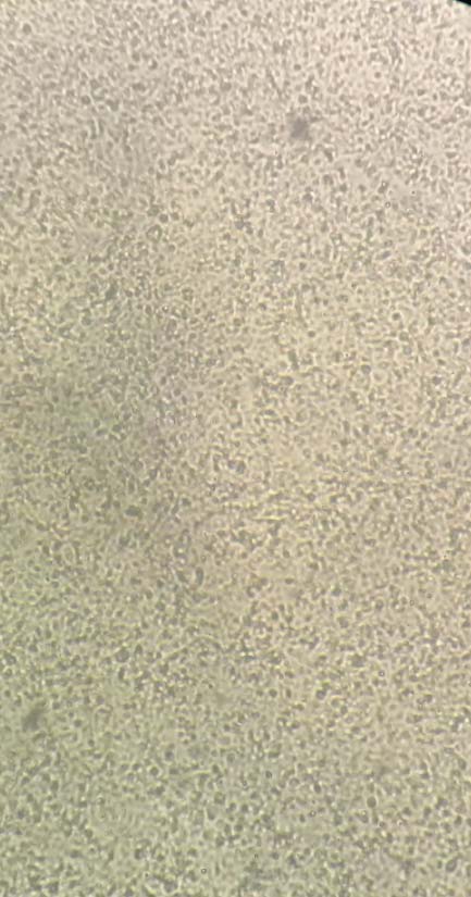
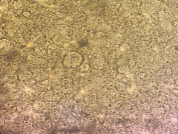
Figure (3) demonstrated a significant growth rate in either MDBK or Vero cells up to 7-10% (EPL) supported cell growth and maintained Viabilities comparable or similar to (NBCS). Significant proliferation achieved at 10% in GS, EPL and NBCS.
Table 2 revealed that cell counting and viability of Vero After 24hr cultured in media containing at 8% EPL, NBCS, and GS were 39, 30 and 31 mean while at 10% were 52.2, 47.8
9. Discussion
Culture of animal cells is key operation in bioscience whether it is related to research at universities or industrial production of pharmaceuticals with the help of gene technology (Hodgson, 1995). Fetal bovine serum (FBS) usually considered as the standard gold serum as a supplement for growth of cell lines. Considerable efforts have been made to reduce or eliminate serum proteins or even using its alternatives as newly born calf serum (NBCS), goat serum (GS) and Equine platelets lysate (EPL) as a serum-free media.
In this work, the first plan was establishing another alternatives to fetal bovine serum by using (NBCS) and (GS). Medium supplemented with (GS) could be used in the propagation of Vero & MDBK cell lines. Blind 7 and 4 successive passages were applied in Vero& MDBK cell lines respectively. figure (1) revealed that cells have been proliferated using goat serum alone compared to GS beside EPL or NBCS (figure, 2) cell growth was achieved in media supplemented with (GS) at concentration 5% & 6% up to 10% in MDBK and Vero cells. Another point of view, the percentage of (GS) could be decreased when 0.5-1% (EPL) was added to growth media. This also achieved a preferable cellular morphology and proliferation rate. The growth rate was higher in GS than NBCS at similar concentration up to 5-8% thereafter become the same. All growth promoting factors were significantly increased at 10%. This finding coincidence with the result of Paranjape, 2004 that 10 % goat serum containing media is similar to fetal serum in many cell line and primary cells except BHK-21 cell line. The author found the relation between DNA, total protein and cell count of Vero cells propagated by using 10% (GS).
Furthermore, the author added that Vero cells adapted & maintained on (GS) were used in preparation of CFT & detection of Dengue virus antigen from clinical samples & interferon production in LM & MFS cells.
The second plan was designated to find alternative use to FBS by (EPL) as a serum free-media .the results in (figure , 3) demonstrated significant growth rate in either MDBK and Vero cells up to 7-10%. The proliferation rate of EPL was nearly similar to NBCS proliferation and cellular viability is dose dependent.
The number of cells correlated with (EPL) % in the supplemented medium that agree with Liselott et al., 2003, concluded that 10% EPL induces cell growth, viability and product formation using a number of target cells including myelomas, hybridomas, hepatocytes, fibroblasts and epithelial cells.
Also Doucet et al., 2005and Horn et al., 2010 have been used (PL) as an FBS substitute in expansion medium (EM) to support the growth of human mesenchymal cells that induced higher mitogenic effect than fetal bovine serum (FBS).
Concerning with the counting and viability of adherent Vero cells after 24hr incubation at 37 0 C using (GS), NBCS and (EPL) in table 2, observed that (EPL) supplemented media induce the best viability for cell culture. There was no apparent difference in Viability between (NBCS) and (GS) in spite of higher died cells in (NBCS) supplemented media.
The result disagrees with Russell & Koch, 2015, recorded no significant difference between the pooled (EPL) and (FBS) treatments in mesencymal (MSC) cells up to a concentration of 30%.
V.
10. Conclusion
A useful alternative to fetal bovine serum in the medium for animal cell culture as a growth-promoting factor could be achieved by using:

| Cell line | of Growth Concentration | Affected wells/ Total No. | |
| factors % | Goat | Platelets | |
| serum | lysate | ||
| 4 | 3/3 | 1/3 | |
| 6 | 0/3 | 0/3 | |
| Vero and | 8 | 0/3 | 0/3 |
| MDBK | |||
| 10 | 0/3 | 0/3 | |
| 12 | 0/3 | 0/3 | |
| 15 | 0/3 | 0/3 | |
| *No. of wells showed microscopically deviation in cells/total | |||
| No. | |||
| Used % | Vero | MDBK | |
| 5 | 30 | 50 | |
| 6 | 50 | 60 | |
| Newly born calf | 7 | 60 | 80 |
| serum (NBCS) | 8 | 80 | 90 |
| 9 | 90 | 100 | |
| 10 | 100 | 100 | |
| 5 | 30 | 30 | |
| 6 | 50 | 60 | |
| Equine platelet | 7 | 60 | 80 |
| lysate (EPL) | 8 | 80 | 90 |
| 9 | 90 | 100 | |
| 10 | 100 | 100 |
| Growth promoting factors | Used % | Viable cells/1ml | died cells/1ml | Total No. | Viability % |
| 5 | 2.35 x10 4 | 1.35 x10 5 | 1.58 x10 5 | 14.8 | |
| Equine platelet lysate | 6 | 9.85 x10 5 | 3.18 x10 5 | 5.12 x10 6 | 19 |
| (EPL) | 8 | 2.05 x10 6 | 4.13 x10 5 | 5.13 x10 6 | 39 |
| 10 | 3.88 x10 6 | 3.54 x10 6 | 7.42 x10 6 | 52.2 | |
| Newly born calf | 5 | 2.3 x10 4 | 1.78 x10 5 | 4.69 x10 4 | 12.9 |
| Serum | 8 | 2.05x10 6 | 6.87 x10 6 | 5.78 x10 6 | 30.2 |
| ( NBCS) | 10 | 3.79 x10 6 | 7.92 x10 6 | 6.90 x10 6 | 47.8 |
| 5 | 2.8 x10 3 | 1.85 x10 5 | 2.16x10 5 | 13 | |
| Goat serum(GS) | 8 | 2.13 x10 6 | 4.05 x10 6 | 5.98 x10 6 | 31 |
| 10 | 3.99 x10 6 | 4.38 x10 6 | 7.57 x10 6 | 47.6 | |
| IV. |