1. Introduction
Author ? ? ?: Department of Pathology, Varhman Mahavir Medical college and Safdarjung Hospital, New Delhi, India. e-mail: [email protected] Introduction cular surface squamous neoplasia (OSSN) is a wide spectrum of disease ranging from mild dysplasia to carcinoma in situ to invasive squamous cell carcinoma of the ocular surface. Most common site is limbus where most actrive mitotic cell reside . [4] OSSN is more common in equatorial countries where exposure to sunlight is more common. Risk factors for OSSN include male gender, ultraviolet light exposure, human papilloma virus (HPV) infection, human immune deficiency virus (HIV) infection. [4], [5], [6], [7] Clinically, the diagnosis is difficult due to variable presentation suspected by the appearance of epithelial changes of the of the ocular surface. OSSN may dissimulate chronic conjunctivitis, corneal ulcer, pterygium, necrotizing scleritis. [8] Slit-lamp examination shows gelatinous, papilliform or nodular lesions. Nodular is being the most common amongst it. [9] Histopathological evaluation is important for the definitive diagnosis. Immuno histochemically detectable p53 protein, bcl-2 protein, MIB-1 are being used as markers of proliferative potency having a possible O prognostic value. [10] Capillary hemangioma is common in pediatric and adult age group and presents in early life. [11] We report a case of OSSN with hemangioma in a 33-year-old male who has been clinically diagnosed with hemangioma and subsequently been treated for it.
2. II.
3. Case Report
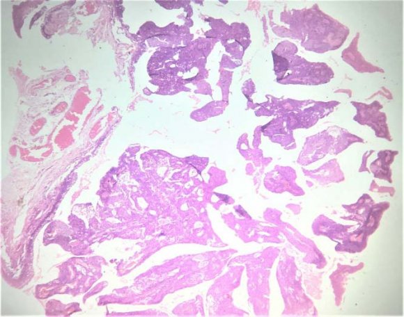
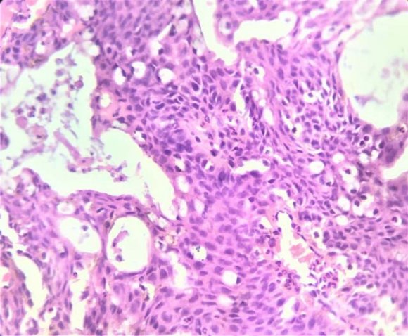
A 33-year-old male presented with complaints of redness associated with itching in left eye for the past 6 month which was intermittent, recurrent and not associated with any painor discharge, eye surgery or trauma. He was a shopkeeper by profession. There was a history of multiple treatment regimens, including artificial tears, topical nonsteroidal anti-inflammatory drugs. He had no systemic illness and there was no lymphadenopathy. On examination, his best-corrected visual acuity was 20/20 both eye. On slit lamp examination of the left eye,a sessile papillary vascular mass in fornix was seen. Engorged conjunctival vessels were seen at the base of the lesion. The surface of the lesion was irregular. Blood investigations were normal. He underwent uneventful surgery in the left eye under local anesthesia .The excised lesion was sent for histopathological examination. Histopathology report showed a columnar lined epithelium of conjunctiva which shows dysplasia along with epithelomatous hyperplasia and papillomatosis. Dysplasia was from moderate to severe degree. Subepithelial tissue showed hemangioma so the diagnosis of ocular surface squamous neoplasia with severe dysplasia and subepithelial hemangioma was made. Postoperatively, the patient was given a course of topical antibioticsteroid combination in tapering dose over 3 weeks, topical lubricants for 6 weeks.
Abstract-OSSN represents the spectrum of disorder ranging from dysplastic changes to squamous cell carcinoma involving surface of an eye. [1] Conjunctival hemangioma is proliferation of blood vessels within the substantia propria. Ocular surface squamous neoplasia (OSSN) is a disease of the elderly [2] in western countries. However in African and certain part of asia, OSSN afflict youger patient and tend to be more clinically aggressive. [1], [3] We report a rare case of a OSSN with hemangioma in a young 33 year old male presented with fornix nodule in left eye. Diagnosis of hemangioma was made based on clinical suspicion. Definitive diagnosis needs histopathological evaluation.
4. Discussion
OSSN is a localized, slow growing lesion with low metastatic potential. OSSN has intraocular and orbital extension rate of 4%, regional and distant metastasis rate of 1.2%, and a mortality rate of 0.8%. Sites of metastasis are preauricular, cervical lymph nodes, the parotid gland, lungs, and bones. Regional lymph node involvement precedes the development of distant metastases. [12] What makes this case interesting is a rare presentation of subconjunctival hemangioma along with ocular surface squamous neoplasia The subconjunctival hemangiomas are rare vascular conjunctival presentation with OSSN is rarer. Symptoms range from none to visual loss. Clinically its difficult to made diagnosis therefore definitive diagnosis is made by histopathological examination of the excision biopsyspecimen. Histopathological diagnosis is vital for both defining therapeutic options as well as for prognostication. [13] Nonsurgical options for small OSSN lesions include topical 5-FU and MMC drops. They eliminate the need to worry about the excised margins. [14] Surgical excision with ''wide margin, no touch'' technique is currently the best established form of treatment. Nevertheless, recurrences of these lesions are common after surgical excision, depending on the involvement of the surgical margins. Recurrence rates following excision of OSSN alone range from 15 to 52%, with an average of 30%. Recurrence rate is 5% when the surgical margins are free and 53% when the surgical margins are involved. [15] In the management of OSSN, excision biopsy, though considered the gold standard. [4] Simple surgical excision is an option in small tumors, but the recurrence rates can be as high as 33% even in tumors excised with clear margins With adjuvant cryotherapy, the recurrence goes down to 12% in tumors excised with clear margins. [16] Surgical excision with adjuvant chemotherapy is another described technique with antimetabolites being given either intraoperatively or as drops postoperatively. [17] In our case, we used a combination of both intraoperative and postoperative MMC.
IV.
5. Conclusion
Slow growth of lesions of OSSN and the ever present malignant potential makes regular follow-up of these patients for the remainder of their life mandatory. Funding: No funding sources Conflict of interest: None declared Ethical approval: Not required