1. Introduction
idney cancer makes up 2% to 3% of adult malignancies, with an incidence of 7 to 10 cases per 100,000 inhabitants in the most developed regions of Brazil [123]. In these cases, clear cell carcinoma represents 81%, followed by papillary carcinoma, which is responsible for about 14% of kidney cancer cases [4]. It is estimated that about 16% of patients are diagnosed with metastatic disease [5]. Cardiac and pericardial metastasis is rare [4]. In the period from 1935 to 1998, there were only 131 cases of cardiac tamponade as an initial manifestation of underlying malignancy [6]. In this case, the patient described in this report developed cardiac tamponade, an event that is triggered by a pericardial effusion. It is a condition that needs urgent intervention. Among the causes, metastatic spread of malignant diseases is an uncommon cause, being clinically silent in most cases [7]. In rarer situations, the clinical presentation can be present, be variable and delay the patient's diagnosis, which is essential to be performed early to reduce morbidity and mortality [8]. We report a case of cardiac tamponade caused by metastasis of a clear cell renal tumor and discussed the symptoms, diagnostic methods and the need for urgent surgical intervention through thoracoscopy, pericardiectomy and making a pericardial window.
2. II.
3. Case Report
Male patient, 49 years old, admitted to Hospital Alberto Cavalcanti in Belo Horizonte -MG, Brazil, on 04/23/2020 with dyspnea and cough started 6 months ago, associated with weight loss of 5kg. He sought care due to persistence and worsening of symptoms, presenting on admission: orthopnea, paroxysmal nocturnal dyspnea, dyspnea on exertion and cough that was accentuated in the supine position. Without fever, sputum or flu-like symptoms, a previous diagnosis of COPD of undetermined origin had already been made, and heart failure was questioned. During the physical examination, he noticed the presence of a mass in the left hypochondrium and lymph node enlargement in the left cervical chain. Computed tomography of the chest and abdomen showing a mass in the left kidney of 15 cm, suggestive of primary neoplasia. Mediastinal lymph node enlargement, bilateral micro pulmonary nodules and 2.7 cm adrenal mass, suggestive of secondary neoplasia (figure 1) Immunohistochemistry showed metastatic clear cell carcinoma, a positive study for cytokeratin and diffuse expression for PAX8. The findings favor that the kidney is the site of origin for this metastatic carcinoma.
To rule out heart failure, a transthoracic echocardiogram with preserved systolic and diastolic function and normal BNPs was performed. Since 05/17, the patient has presented progressive worsening of the respiratory pattern and refractoriness to clinical treatment. On 05/24, the patient was evaluated in bed, with respiratory worsening, tachycardia, jugular engorgement, paradoxical pulse, pain in the right hypochondrium, orthopnea, auscultation with expiratory wheezing and light crackling. Urgent ultrasound was performed, identifying pericardial effusion and indirect signs of cardiac tamponade (Figure 2). Thoracic surgery was requested to be evaluated due to the patient's instability, and he was immediately referred for a surgical approach, where thoracoscopy was performed. 10 cm incision in the region of the 6th left intercostal space with 10mm trocars and 30mm optic passages within the left hemithorax inventory. Engorged pericardial effusion was identified with subsequent opening of the pericardium with a Hook cautery pen and elimination of bloody liquid (Figure 3). Pericardial window and pericardiectomy of about 2 cm² were performed (Figure 4). Approximately 200ml of hematic pericardial content was aspirated, and there was an instant improvement in hemodynamic parameters. Pericardial material and pericardial implant were collected for biopsy. Finally, water seal pleural drainage was performed. Anatomopathological examination of the pericardial biopsy confirmed metastatic implantation. There were no complications in the postoperative period, a chest tube was removed on the 12th postoperative day, and the patient was discharged on 06/16/2020, referred to the clinical oncology clinic for therapeutic follow-up.
4. Discussion
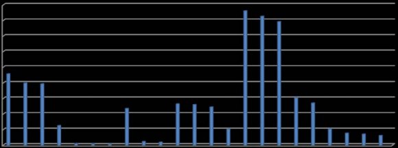
Malignant tumors represent 32% of the causes of cardiac tamponade [9]. Pericardial metastases have been reported in 15.4% of cases of malignant autopsied tumors, but most are asymptomatic and rarely cause clinical repercussions [10]. According to Oliver et al. [11], the main primary malignant tumors with pericardial metastasis are lung cancer 36.5%, breast cancer 22.3%, leukemia and malignant lymphoma 17.2% and renal cancer 1.9%. As a metastatic route for pericardial metastases, it is speculated that it is done by retrograde lymphatic transit [12].
In the period from 1935 to 1998, there were only 131 cases of cardiac tamponade as an initial manifestation of underlying malignancy. The main primary cancers causing effusion were: lung (52 cases), lymphomas and leukemias (17 and 19 cases respectively). Only one case of renal cell cancer was reported during this period [6]. A study carried out between January 1, 1999 and January 31, 2003 evaluated 219 patients with pericardial effusion, 96 patients had the disease related to malignancy, only one positive case for renal cell cytology (table 1). [13] The frequency of cardiac tamponade as the initial manifestation of malignant effusion is highly variable and depends on the rate of fluid accumulation, fluid volume and underlying cardiac function. The pericardium can be stretched for a period to accommodate a large volume of fluid before the clinical appearance of the tamponade. The signs and symptoms of cardiac tamponade include dyspnea, orthopnea, low output (peripheral vasoconstriction, cold and wet extremities, poor capillary filling and diaphoresis), jugular venous distention, muffled heart sounds, paradoxical pulse and reduced pulse pressure. Even with cancerous pericarditis, in the case of kidney cancer, performing pericardiocentesis not only reduces symptoms, improving heart failure, but also prolongs the period of survival. The median overall survival of patients with malignant pericardial effusion is less than 6 months [14].
In the present case report, we narrate a case of clear cell renal cell carcinoma, the first symptoms presented by the patient were cough and dyspnea, although the first echocardiogram did not show significant changes and the patient had a previous COPD to clarify, the treatment established for COPD and HF were not effective. The symptoms presumably were due to the ongoing pericardial effusion. One week after CT showing bilateral pleural effusion and small pericardial effusion, with the exacerbation of cardiac symptoms, ultrasound at the bedside was performed, which showed an important pericardial effusion with cardiac tamponade.
Videoracoscopy and pericardiectomy were indicated by the surgical team. Videothoracoscopy was chosen because it is less invasive than open thoracotomy, the pericardial approach through electrocauterization could be performed by a previous study via POCUS, better viewed in the lower left parasternal window. Approximately 550 ml of hematic pericardial content was aspirated, with instant improvement in hemodynamic parameters.
Although simple pericardiocentesis can save lives in cases of cardiac tamponade, this procedure alone is rarely an adequate therapy due to the high rate of fluid buildup. To avoid recurrence, the patient received immunotherapeutic treatment and pericardiectomy was performed against urgent pericardiocentesis, provided by the shunt in the pericardial window in the case confirmed by the pathology of clear renal cell metastasis. Clear cell renal cell carcinoma that presents as cardiac tamponade is rare in the literature. The case also emphasizes the importance of a complete review of the history, physical examination and complementary examination, in addition to exposing the need to perform a resolutive surgical procedure.

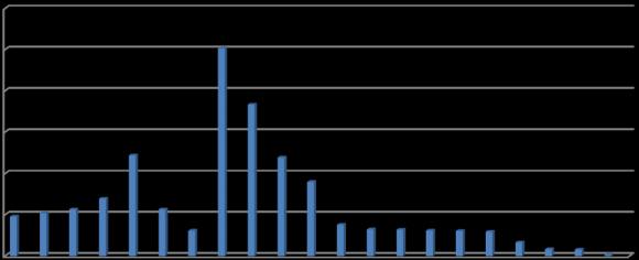
| Patients | ||
| Site | No. % | |
| Lung | 33 | 34.4 |
| Breast | 16 | 16.7 |
| Leukemia/myelodysplastic syndrome | 9 | 9.4 |
| Cancer of unknown primary | 8 | 8.3 |
| Esophagus | 5 | 5.2 |
| Sarcoma | 5 | 5.2 |
| Mesothelioma | 4 | 4.2 |
| Lymphoma/lymphoproliferative disorder | 4 | 4.2 |
| Colorectal | 4 | 4.2 |
| Other* | 8 | 8.3 |
| Total | 96 | 100.0 |
| *Other malignancies: cervical (2), head/neck, eccrine gland, nerve sheath, ovarian, prostate, renal cell. [8] | ||