1.
Post-Traumatic Duodenal Stenosis following a Duodenal Hematoma: Case Report and Review of the Literature Introduction ost-traumatic duodenal hematomas are rare and pose a diagnostic problem because of retropancreatic situation of the duodenum. 70% of duodenal hematomas occur after blunt abdominal trauma (1). The first case of duodenal hematoma was published in 1838 by McLaughlan who described it as "a fatal pseudoanevrysmatic swelling" (2) Early diagnosis and adequate therapy are essential because a delay beyond 24 hours, increases mortality by 11 to 40%. Retroperitoneal attachment and the absence of mesentery, as well as the proximity of the horizontal part of the spine, mayexplain the vulnerability of the duodenum to blunt abdominal trauma (3), the clinical signs are non-specific and vary according to the site of the hematoma in the four portions of the duodenum. We report the case of a patient victim of a public road accident causing a blunt abdominal trauma manifested by vomiting and abdominal pain .the abdominal CT showed the presence of a duodenal hematoma compressing the 3rd duodenal portion responsible for gastric distension upstream treated by a conservative method.
Author ?: Visceral Surgical Emergency Department, Faculty of Medicine and Pharmacy, University Hospital Center Ibn Rochd, Hassan II University, Casablanca, Morocco. e-mail: [email protected] II.
2. Patient and Observation
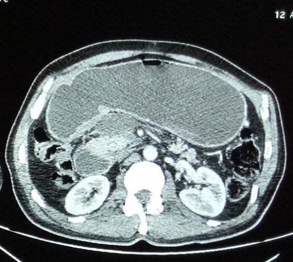
A 55-year-old male patient, diabetic under oral antidiabetic drugs, a victim of a public road accident causing a blunt abdominal trauma, who presented five days after his accident, an early post prandial then bilious food vomiting associated with epigastralgia, on clinical examination, presence of epigastric tenderness. A bodyscan was requested showing a collection attached to the anterior wall of the 3rd portion of the duodenum compressing its lumen giving an important gastric, duodenal and esophageal stasis measuring 49*19 mm (figure.1).)
CT scan showing a duodenal hematoma (red arrow) responsible for a stasis stomach (blue arrow) A biologic check-up was requested showing hypokalemia at 2.7 mmol/ associated with functional renal failure with urea at 1.3 g/l and creatinemia at 40 mg/l; a blood count showed hemoglobin at 10 g/dl, platelet count was normal at 320*103 /mm3, hemostasis was correct and lipasemia was normal. Management was conservative by monitoring and conditioning the patient with a rehydration regimen to correct electrolyte disorder; a nasogastric tube was performed to aspirate gastric secretions and stop vomiting, and parenteral nutrition was instituted. After 15 of clinical and biological surveillance we observed the improvement of the patient by drying up of the vomiting and correction of the biological balance sheet, the control CT scan was normal.
3. III.
4. Discussion
Duodenal lesions after blunt abdominal trauma are 3-5%(4), more frequent in men (80%) (1). The duodenum has no mesentery and can be divided into four parts, the first, upper, part is located intraperitoneally, while the three distal parts are located retroperitoneally. The most affected duodenal portion is the second (36%), followed by the third (18%) and fourth (15%), while the least common sites for duodenallesions are the first (13%) and multiple proportion lesions (18%) (5). The retroperitoneal attachment and the absence of mesentery, as well as the proximity of the horizontal part with the spine, mayexplain the vulnerability of closed abdominal trauma (6) The suspension of the duodeno-jejunal junction at the level of the Treitz ligament is also considered as a privileged site of post-traumatic duodenal hematoma (7) duodenal hematoma can occur following several causes outside a traumatic context, notably during a duodenal ulcer, pancreatitis and iatrogenic causes such as duodenal biopsy, after fibrodoscopy or after anticoagulant overdose (3) clinical signs are highly variable and nonspecific and vary according to the site of the hematoma in the four duodenal portions and are represented by signs of duodenal obstruction manifested by early and late post-prandial vomiting, Epigastralgia, when the hematoma compresses the papilla of Vater causes cholestasis or even pancreatitis, Patients with significant intramural hematomas are at risk of developing anemia or even hypovolemic shock (8). Biological signs are non-specific except for a decrease in hemoglobin levels associated or not with an elevation of pancreatic or hepatic enzymes (1). abdominal CT scan with ingestion of contract productis the standard gold test for a positive diagnosis, endoscopy and MRI are requested in case of diagnostic doubt(9) In a 6-year study by Ballard et al, abdominal CT scan made the diagnosis in 40% of cases (10) initially the treatment of duodenal haematomas was essentially surgical, ranging from incision and surgical drainage of haematomas to the insertion of a gastrojejunostomy followed or not by a bypass (11) currently the management has completely changed towards a more conservative strategy by reserving surgery for persistent occlusions and expansion of the hematoma (12) surgery is urgently required in case of suspicion of perforation where as it could be delayed and performed after 7-14 days if there is no improvement (1). Drainage should be considered before any laparotomy, evacuation of the hematoma can be performed by CT or ultrasound guided procedures. It may be performed even endoscopically (13). Arterial embolization can be used to stop bleeding. Conservative treatment, including gastric decompression, parenteral nutrition and antibiotic prophylaxis, may be chosen (11). The results of conservative treatment are favourable with complete resolution of duodenal hematoma within 2-3 weeks (13).
IV.
5. Conclusion
Duodenal hematoma is an acute disease whose possible complications can be prevented by early diagnosis, these complications range from obstruction I or narrowing of the duodenum to pancreatitis, or even hemorrhage by erosion of the hematoma which can lead to hemorrhagic shock. Diagnosisis based on CT scan and treatment is essentially conservative, surgery may be necessary if conservative methods fail.
6. Authors' contributions
This work was carried out in collaboration among all authors. All authors contributed to the conduct of this work. They also declare that they have read and approved the final version of the manuscript.
7. Consent
According to the international or academic standard, patient consent was collected and retained by the authors.
8. Ethical Approval
As per international standard written ethical approval has been collected and preserved by the author(s).
9. Competing Interests
Authors have declared that no competing interests exist.