1. Background
aginal metastases in patients with clear cell renal carcinoma are rare. There are fewer than 100 cases currently described as the revised literature. At the time of diagnosis, we observe metastasis by hematogenous or lymphatic spread in 20-30% of patients [1].
Vaginal metastases, despite being rare, are more common than primary tumors and as presentation of the disease is extremely rare.
We present the evolution and treatment of a patient with renal cell carcinoma and vaginal metastasis.
2. II.
3. Case Presentation
We report the case of 68 years old caucasian female, with a history of hypertension, Sjögren's syndrome, vitamin D deficiency, mild mitral regurgitation and mild aortic regurgitation.
The patient has constitutional syndrome associated with macroscopic hematuria for 4 months evolution.
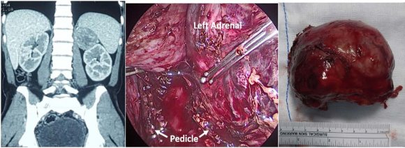
On physical examination we find a palpable mass in the left flank and evidenced gross hematuria. We performed a pelvic abdominal CT scan that evidence a heterogeneous mass of 10 x 16 x 9 cm dependent on the back side, the middle and lower third of the left kidney. Also striking, the presence of bilateral pulmonary parenchymal involvement with multiple nodular formations compatible with mestastasica involvement (the greater than 4 cm) and apparent hilar and mediastinal infracarinal lymph nodes. The clinical stage was cT3 No M1. This case was discussed in the uro-oncology committee and despite being classified as intermediate risk according to MSKCC/Motzer's criteria [2], the patient was intervened because she had symptomatic disease.
The patient underwent a left laparoscopic radical nephrectomy.
The study of the specimen reveals a mass of 9x6x8 cm located in the middle third and lower pole, infiltrating the renal capsule, the renal sinus, perirenal fat, renal vein and numerous segmental renal veins. In addition, numerous tumor thrombosis in lymphatic vessels of renal sinus were observed (Figure 1).
Regarding lymph node staging, lymph node metastases were seen in one of the two extracted hilar lymph and in two of the three nodes obtained from the specimen of regional lymphadenectomy. On microscopic analysis we observed a renal clear cell carcinoma, grade 4 (ISUP 2014/WHO 2016) in 90% of the tumor, Fuhrman IV (90%), with 40% tumor necrosis with negative surgical margins. The pathological stage was pT3a N2.
In the first postoperative day, the patient has an important episode of metrorragia. On genital examination we found a solid mass of 2x2 cm in the left lateral surface of the vagina, on the lower third. Abstract-Background: Vaginal metastasis, despite being rare, are more common than primary tumors and as presentation of the disease is extremely rare. At the time of diagnosis metastasis by hematogenous or lymphatic spread in 20-30% of patients.
Case presentation: 68 years old female patient that debuted with haematuria. In the extension study we can objectify a left renal mass treated by laparoscopic radical nephrectomy.
During admission the patient presented and episode of metrorragia. A lesion was found in the lower thrid of the vagina, which was biopsed, resulting a vaginal metastasis of clear cell carcinoma. The patient presented a favorable evolution being discharged four days after de surgical intervention. The subsequent extension study revealed progression of the underlying disease with mediastinal nodes and bone metastases.
The patient died 9 months after surgery having received treatment with tyrosine-kinase inhibitors.
Conclusions: About 30% of patients diagnosed with renal carcinoma have metastases at the time of diagnosis. Vaginal location is extremely rare and usually occurs with episodes of metrorrhagia and mass effect. Treatment consists on removal of the lesion or local radiotherapy. The prognosis of these patients is conditioned by metastases in other organs.
The patient underwent emergency surgery to suture the vaginal tear and excisional biopsy of the lesions.
The pathology of both lesions revealed metástasis of clear cell renal cell carcinoma with high grade nuclear atypia and abundant lymphovascular tumor thrombosis ( Figure3 and 4). The patient had a favorable evolution getting discharged from hospital four days after nephrectomy.
One month after discharge the patient begins treatment with pazopanib (800 mg / day). A thoracoabdomino-pelvic CT control done two months after surgery reveals an acute pulmonary embolism on lobar artery on right lower lobe as well as progression of the underlying disease with increased number and size of hiliar lymphnodes and mediastinal conglomerates adenopathicas well as lytic lesion in the posterior arch of the 5th costal left rib with probable metastasic origin. The patient died 9 months after surgery.
4. III.
5. Discussion and Conclusions
Since the 1970s the incidence of RCC has increased given the use of ultrasound and CT routinely for the diagnosis and evaluation of various abdominal disorders.
The classic triad of hematuria, flank pain and palpable mass is observed between 5% and 15% of patients and may also debut as different paraneoplastic syndromes like Cushing's syndrome, Stauffer syndrome, deep vein thrombosis or amyloidosis among others.
At the time of diagnosis, we can observe metastasis by hematogenous or lymphatic spread in 20-30% of patients [1]. The most frequent locations are retroperitoneal nodes, lung, liver and bone.
Vaginal adenocarcinomas are rare entities (5% vaginal cancers) and almost always metastatic (91%). In young women, they are often related with exposure to diethylstilbestrol. In older women, as in our case, they are almost always metastatic.
The appearance of the vaginal lesion usually precedes the diagnosis of the primary tumor, and the presenting symptoms, usually metrorrhagia, vaginal discharge and mass effect.
Vaginal metastases are more common in tumors located in the left kidney and generally, these metastases occur ipsilateral to the primary kidney tumor. Less than 90 cases of vaginal metastasis of RCC were reported. In most of these cases, vaginal metastases were diagnosed as metachronous metastatic disease that discovered long term after radical nephrectomy. There are only four cases of synchronous vulvo-vaginal metastases from RCC in medical literature [3][4][5][6].
As for the way of disemnicacion, JJ Mulcahy proposed the theory that even today remains the most plausible explanation for this phenomenon [7].
The spread is caused by retrograde venous extension from the renal vein to the ovarian vein, ovarian plexus and uterovaginal plexus [8].
The most important prognostic factor in these patients is the presence or absence of metastases in other organs. No conclusive studies in the literature that support the realization of Pap smear for the diagnosis or monitoring of these patients.
The treatment of metastatic renal cancer has varied over the last decade. Currently, target therapies such as tyrosine kinase inhibitors and immunotherapy are used depending on the risk of the disease. Cytoreductive nephrectomy is reserved for those patients with a more favorable prognosis or those with symptomatic disease.
As therapeutic recommendations premature ovarian vein ligation during nephrectomy it is recommended in order to limit the migration of tumor cells from the renal vein [9].
Local excision and / or radiotherapy are the standard treatments for vaginal metastases in patients undergoing radical nephrectomy [10].
About 30% of patients diagnosed with renal carcinoma have metastases at the time of diagnosis. Vaginal location is extremely rare and usually occurs with episodes of metrorrhagia and mass effect. Treatment consists on removal of the lesion or local radiotherapy. The prognosis of these patients is conditioned by metastases in other organs.
6. List of Abbreviations
Abbreviations: RCC, Renal cell carcinoma; CT, Computerized tomography; MSKCC, Memorial Sloan Kettering Cancer Center, ISUP, International Society of Urological Pathology; WHO, World Health Organization.
7. Declarations
Ethics approval and consent to participate: The hospital ethics committee endorsed the review of this case. Hospital La paz Ethics Comitte.
8. Availability of data and material:
The data was collected from the electronic medical record of the hospital The fact that approximately 60% of RCC with vaginal metastases are located on the left side favors this hypothesis.
analysis of the specimen. All authors read and approved the final manuscript.