1. Introduction
utembacher Syndrome (LS) is a rare cardiac clinical entity characterized by the unusual combination of atrial septal defect (ASD) and Mitral stenosis (MS) (commonly of rheumatic nature) [1].
In the first description of LS by the frensh physician Rene Lutembacher in 1916, after whom this syndrome was eventually named [2], both lesions were thought to be congenital in origin, but since then, the definition has been broadened.
In a typical LS case, the ASD is usually more than 15 mm in size, which can cause progressive pulmonary hypertension (PH) [3], however, in the current era of percutaneous balloon mitral valvuloplasty (BMV) for acquired MS, ASD can be secondary to transseptal puncture, which defines a new entity called iatrogenic LS [4].Thus, the current consensus defines LS as any combination of ASD (congenital or iatrogenic) and MS (congenital or acquired) [5].
Clinically the syndrome may resemble either isolated ASD or MS, depending upon the dominant lesion [6], but can also present in unusual forms.
LS is generally associated with long-term unfavorable natural course [7] depending on the evolution of PH and the occurance of heart failure (HF).
Author ? ? ? ? ¥ §: Department of cardiology, Hospital University Center Ibn Rochd, Casablanca, Morocco. e-mail: [email protected] Surgical and percutaneous trans-catheter therapies have proven to be beneficial in patients with LS [8].
We here report the case of a young male who was diagnosed with Iatrogenic LS based on relevant clinical findings and investigations, stressing the major role of echocardiography in the contemporary diagnostic and therapeutic modalities for LS, which can be challenging in the context of developped countries.
2. II.
3. Case Presentation
We report the case of a 46-year-old male with history of rheumatic mitral stenosis (MS) for which he successfully underwent percutaneous transvenous mitral commissurotomy (PTMC) 7 years ago, who presented to the emergency department (ED) with gradually progressive exertional dyspnea, fatigue and palpitations.
Physical examination found a blood pressure of 99/64 mmHg, an irregular pulse rate of 61/min in addition to a tapping apex impulse, jugular vein distention, bilateral ankle oedema and abdominal distention suggesting ascites.
On cardiac auscultation, the first heart sound was loud, the second heart sound was widely split with a prominent pulmonary component. He also had a grade 4/6 diastolic rumble increasing with expiration in the apex, associated with a grade 2/6 holosystolic murmur and a grade 3/6 pansystolic murmur in the tricuspid area. Respiratory examination revealed bilaterally equal normal breath sounds with endinspiratory fine crackles at the base of both lung. Electrocardiogram (ECG) showed right-axis deviation and atrial fibrillation (AF) (Figure 1). Chest radiograph showed cardiomegaly with a cardiothoracic ratio of 0.61, pulmonary plethora with biatrial and right ventricular enlargement.
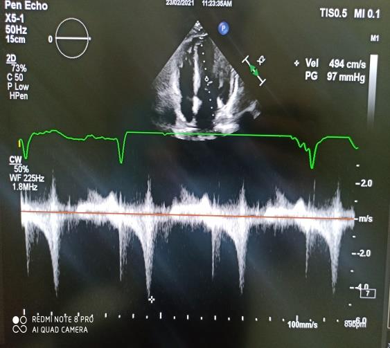
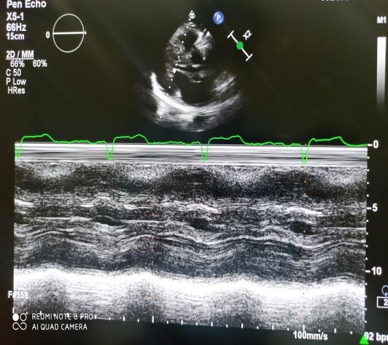
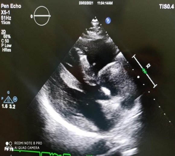
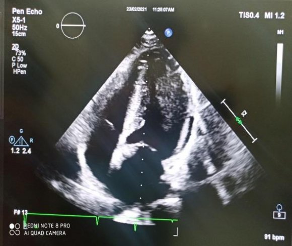
Transthoracic echocardiography (TTE) revealed calcified and thickened mitral valve leaflets with bicommissural calcification and a "hockey stick" appearance of anterior mitral leaflet and immobility of the posterior leaflet with severe mitral stenosis (MS)(Mitral Valve Area (MVA) was 0.5 cm² by planimetry and mean pressure gradient (MPG) was 18 mmHg) associated with amoderatemitral regurgitation (MR) (Figure 2). The Aortic cusps were also thick and calcified with a moderate aortic stenosis (AS) with a MPG of 18 mmHg and an aortic valve area of 1,1 cm2, associated to a mild aortic regurgitation (AR). Left ventricular (LV) function was normal, with a Left ventricular ejection fraction (LVEF) of 57% and a global longitudinal strain (GLS) of -19.1%. LV dimension during systole (LVDs = 26cm/m²) and diastole (LVDd = 31cm/m²) were normal, Interventricular septal thickness (IVST) and Posterior wall thickness (PWT) were also normal (7 and 8 mm respectively). The right ventricule (RV) was dilated (RV basal diameter of 45 mm) with a preserved function and a severe functional tricuspid regurgitation (TR) with an estimated Pulmonary artery pressure of 75 mmHg and a dilated inferior vena cava. In addition to these findings, we assessed bi-atrial enlargement with a left atrial spontaneous echo contrast and a non-restrictive Atrial Septal Defect (ASD) of the ostiumsecundum variety measuring 11mm with bidirectional shunt (Figure 3). The was found in the last TTE after his percutaneous intervention, thus, the diagnosis of iatrogenic Lutembacher syndrome (LS) was suspected. The Transesophageal Echocardiography (TEE) confirmed the diagnosis by showing left atrial spontaneous echo contrast and a non-restrictive ASD with bidirectional shunting in addition to the severe MS(Figure4).
We initiated medical therapy in our patient with high dose furosemide (250mg/Day), Acenocoumarol 4mg and Digoxin 0.25mg/Day. The case was then discussed by the heart-team who decided to perform a double valve replacement and a tricuspid annuloplasty given the associated aortic disease and the severe TR, in addition to a surgical closure of his ASD using a pericardial patch.
In time of submission, the patient successfully underwent surgery with good clinical evolution.
4. III.
5. Discussion
The association of ASD with various abnormalities of the mitral valve (MV) such as MV systolic prolapse with MR are frequently reported, however, MS coexisting with ASD remains rare and represents a distinctive syndrome going by the name of its inventor Lutembacher [2]. At first, the ASD and MS in LS were both thought to be congenital, but since then, the definition has changed several times [5]. The ASD may be congenital (ostiumsecundum or sinus venosus type) or iatrogenic, secondary to cardiac interventional procedures like mitral valvuloplasty [9], as it was the case of our patient. The MS is often acquired in this syndrome as a consequence of rheumatic heart disease (RHD), especially in developped countries, but may also be congenital in rare cases (accounting for only 0.6% of congenital heart disease) [5,7].
The exact prevalence of LS is not well known [10], since it is more prevalent in developped countries with RHD, however, it is a rare syndrome occurring in 4-7% of cases of ASD and 0.6-1.2% of cases of MS [6,7]. LS can present at any age but is usually more common in young adults in the third decade with a predilection for females [7] unlike our patient who was a male which was described only once in the available literature [11].
LS is particular due to its unique haemodynamic consequences that result from the interplay between the ASD and MS. The clinical scenario of this syndrome depends upon the MS severity, the ASD size, the RV compliance and the pulmonary vascular resistance [7].
The third scenario is our patient's case with both severe MS and non-restrictive ASD. Due to the RV better compliance, the blood shunts through the ASD instead of backing up into the pulmonary veins, allowing the Left atrium (LA) to decompress in the right atrium (RA), thus avoiding the rise of left a trial pressure in proportion to the MS severity. Therefore, LS is well tolerated by patients for a long time and pulmonary congestion usually doesn't occur until late in the disease. Thus, symptoms of MS such as exertional dyspnoea, orthopnoea and paroxysmal nocturnal dyspnoea are less severe and delayed due to LA decompression [12].
However, this happens at the cost of an increased pulmonary vascular resistance and left to right shunt across the ASD with progressive dilatation of both the RA and the RV leading ultimately to RV failure and decreased systemic cardiac output. Thus, LS patients usually complain from fatigability on ordinary physical exertion and palpitations [13].
Our patient had signs of RV failure and severe tricuspid regurgitation, which is indicative of right ventricular dysfunction. He also complained from exertional dyspnea, and had end-inspiratory lung crackles, with signs of pulmonary vascular congestion in his chest x-ray, further suggesting that the RV compliance has diminished considerably enough to reduce the amount of shunting via the ASD. The natural course of our patient's disease without treatment would be an irreversible pulmonary vascular disease and the development of an Eisenmenger syndrome, which is usually very uncommon and delayed due to the MS. Another instance of development of a right-to-left shunt is a rare entity called Reverse Lutembacher syndrome, in which a severe tricuspid stenosis is associated to the classic LS thus precipitating central cyanosis, digital clubbing and hepatomegaly [14].
Among other complications of LS, the risk of infective endocarditis (IE) is increased by the presence of MS unlike in isolated ASD. However, the Mitral valve is
6. I
The first clinical scenario is when the MS is severe and ASD is restrictive, the shunt across the defect will be less, and hence, the patient will follow the course of isolated MS. In the same way, in the second scenario, if the ASD is non-restrictive and MS is not severe, the symptoms are those of ASD alone. less calcified in LS, because of the LA decompression, resulting in less turbulent flow across the mitral valve [10]. Furthermore, LA enlargement in LS predisposes the patients to develop AF, which explains the palpitations in our patient.
Preoperative diagnosis of LS is sometimes difficult, Steinbrunn et al [6] have emphasized the dangers of incomplete diagnosis before surgery. They reported 3 patients with LS who underwent surgery for closure of "isolated" ASD, resulting in death by severe pulmonary edema in one case and reopening of the shunt in the others during the early postoperative period. The diagnosis of LS is confirmed by Two-dimensional echocardiography [15]. The severity of MS and the size and type of ASD are accurately estimated, transmitral gradient is less despite severe MS, planimetry is the more reliable method to assess the MVA in LS as compared to Doppler half-time which tends to overestimate the calculations [16]. TEE outlines the site and size of ASD with its flow pattern and is usually a superior imaging diagnostic modality than TTE, which is why we performed both in our patient. Cardiac catheterization is rarely required for the diagnosis of LS, except for the assessement of pulmonary artery hypertension reversibility in Eisenmenger Syndrome, measurement of the MVA if the echocardiography lacks precision, and the evaluation of the coronary anatomy in high-risk patients.
As for the treatment, traditionally LS has been treated by open heart surgery by Open mitral valvotomy (OMV) or mitral valve replacement (MVR) with surgical repair of the ASD). But recently, with the advancement of percutaneous interventional techniques, percutaneous trans-catheter therapy in the form of balloon mitral valvuloplasty for MS and Amplatzer atrial septal occlude for ASD, has gained preference over surgery due to its faster recovery time and decreased length of hospital stay [17,18].
However, contraindications of percutaneous intervention include presence of left atrial thrombi, inadequate rim tissue surrounding the atrial septal defect and anomalous pulmonary drainage [4]. Another contraindication and the reason why our patient could not undergo such procedures is the presence of bicommissural calcification. Moreover, our patient also had a previous PTMC in addition to an associated aortic disease and a severe TR, which made the heart team decide to perform a double valve replacement and a tricuspid annuloplasty.
A symptomatic treatment with diuretics to relieve the symptoms of right-sided HF and/or pulmonary venous congestion in addition to Betablockers and calcium channel blockers for rate control in AF, as well as IE prophylaxis, is strongly recommended.
IV.
7. Conclusion
Persistent iatrogenic ASDs have become an increasingly common finding after invasive procedures requiring trans-septal puncture. Iatrogenic LS is a rare yet challenging disease that needs to be diagnosed correctly via transthoracic and transesophageal echocardiograms in order to provide early and adequate medical and surgical therapies, in order to prevent the onset of PH and HF, thus improving survival rates.
Appropriate surgical procedures, be it transcatheter procedures or open heart surgery should be carefully discussed by the heart team, after an overall assessment of disease progression and cardiac anatomy.