1. Effect of Secretome in Discogenic Pain
Abstract-Study design: This work is an analytical and experimental study. Low back pain has become one of the principal leading cause of work disability in recent decades. Stem cells have emerged as a key element in regenerative medicine therapies, thus providing numerous potential cell therapies in the treating of a range of degenerative diseases and traumatic injuries. Stem cells can secrete potent combinations of trophic factors which modulate the environment of molecular composition to evoke responses from resident cells.
Objectives: The aim of this work is to make an attempt at stopping an irreversible inflammatory cascade at some point using adipose tissue mesenchymal stem cells derivedconditioned medium as therapeutic strategy.
Materials and methods: An in vitro model of inflammation of intervertebral disc has been developed in order to evaluate the effectiveness of secretome from the culture of adipose tissue mesenchymal stem cells to produce an immunomodulation of pro-inflammatory cytokines and neurotrophic factors.
Results: Genes analyzed in the in vitro model of discogenic pain showed how their expression developed when annulus fibrosus and nucleus pulposus cells had been stimulated with tumor necrosis factor. Also, the secretome was capable of modulating the response of various pro-inflammatory cytokines produced in the pathogenesis of discogenic pain. Conclusions: Our data showed the immunomodulatory role of mesenchymal stem cells-derived secretome reducing inflammation and these effects could be a key element in inhibiting the activity of the molecular factors involved in the process of tissue degradation and possibly pain control in IVD degeneration pathology.
2. Introduction
tudies have suggested that between 60-90% of population is affected by low back pain, a debilitating disorder which decreases productivity and has caused considerable therapeutic expenses [1]. The intervertebral disc (IVD) is an avascular and aneural cartilaginous structure composed of the central gelatinous nucleus pulposus (NP), the surrounding elastic annulus fibrosus (AF), and the cartilaginous endplates [2]. Degeneration of the intervertebral disc and herniation is associated with back pain [3]. Although their etio-pathogenesis is not fully understood, it is known that a complex interaction between molecular and biomechanical procedures of the spine which may leads to tissue degradation and thus, loss of function and pain in the IVD [4].
Degeneration is thought to be mediated by both the NP and the AF cells as well as macrophages, T cells and neutrophils which induce the abnormal secretion of pro-inflammatory cytokines [5]. These molecules trigger off a scope of pathogenic responses by NP and AF cells which can promote different responses as autophagy, senescence and apoptosis [6,7]. These secreted proinflammatory mediators include Tumour Necrosis Factor (TNF), Interleukins: IL-1 ?/?, IL-6, IL-17, IL-8, IL-2, IL-4, IL-10 and Interferon gamma (IFN-?). TNF is involved in disc herniation and nerve irritation [8,9]. In addition to the infiltration of immune cells in the disc, there is also proliferation of nociceptive nerve fibres derived from the dorsal root ganglion (DRG) [2,10]. At this point, cytokines as well as neurogenic factors particularly Brain-derived Neurotrophic Factor (BDNF) and Nerve Growth Factor (NGF) are released by NP and the infiltrating immune cells [7].
So many treatments are currently being used to treat inflammatory diseases such as IVD disease. However, these treatments are costly, unspecific, and none of them lead to a remission of pain in the absence of medication. Over the last decade, mesenchymal stem cells (MSCs)-based therapy has emerged as a novel treatment and they have been proposed as the most interesting candidate cells when treating IVD degeneration and herniation [11,12]. Multipotent mesenchymal stromal cells or mesenchymal stem cells are one of the promising cell types for the use in regenerative medicine, particularly in the treatment of musculoskeletal disorders [13].
Initially, therapeutic effects of transplanted MSCs were regarded as being mediated by the homing of MSCs to a damaged site and differentiating in the cells concerned. However, several studies have recently indicated that MSCs exert its therapeutic effects by secreting trophic factors [14]. The stem cell secretome, also referred to as conditioned medium (CM), is a collective term for the paracrine soluble factors produced by stem cells. These factors are thought to control angiogenesis, immune response, protection of the tissue and to accelerate wound healing. Hence, if therapeutic effects based on MSCs are replaced with trophic factor(s) derived from MSCs, there would be great advantages [15].
The aim of this work is to study the protective effect of conditioned medium from adipose tissue mesenchymal stem cells (ASCs) in an in vitro proinflammatory model of IVD degeneration. For this purpose we analyse if CM is capable of performing an immunoregulation on cytokines and the factors responsible of IVD inflammation.
3. II.
4. Materials and Methods
5. a) Biological material
Adipose derived-mesenchymal stem cells ASCs, nucleus pulposus (NP) and annulus fibrosus (AF) human cells were commercially obtained from Innoprot®.
6. b) ASCs culture and conditioned media obtaining
ASCs were multiplied in monolayer T150 flasks (Corning®) at a density of 1x10 6 cells/mL using lowglucose DMEMc at 37°C in a humid atmosphere containing 5 % CO 2 . CM was obtained from cells every 48 h of culture at passages 0 and 1, centrifuged at 2,000 g and conserved until their use at ?80 o C.
7. c) Characterization of ASCs
In order to characterize ASCs the expression of specific markers was determined using Flow Cytometry and Confocal Microscopy.
8. d) Flow Cytometry Analysis
With the aim of confirming the identity of ASCs, the expression of different surface markers was determined: anti-CD73, anti-CD90 and anti-CD105 (1:100) (Abcam®). Cells were stained with secondary streptavidin-Alexa 488 antibodies (1:100) (Invitrogen®). In order to conduct cytometry, 1×10 6 cells per sample were used. Three samples and a control (only secondary antibody) were used for each cell type. Cells were acquired in a FACS Cyan ADP (Dako®). About 1x10 4 events (minimum) were used for fluorescence capture with Summit 4.3 (Cell Quest®) software. All the experiments were developed using three biological and three technical replicates.
9. e) Confocal Characterization of ASCs
On the 8-well Nunc Lab-Tek Chamber Slide System® (2x10 3 cells/well) ASCs were sub-cultured. Cells were fixed with 2% paraformaldehyde for 15 minutes. Then they were incubated with primary mouse anti-CD73, CD-90 and anti-CD105 antibodies (1:100) (Abcam®) overnight at 4°C. Samples were treated with secondary biotinylated anti-mouse antibodies (1:100) (Abcam®). They were then stained with streptavidin-Alexa 488 antibodies (1:100) (Invitrogen®). Finally, Vectashield mounting medium containing DAPI was used. All the experiments were developed using three biological and three technical replicates.
10. f) Confocal Characterization of AF and NP cells
The AF and NP cells were sub-cultured on 8well Nunc Lab-Tek Chamber Slide System® (2×10 3 cells/well). Cells were fixed with 2% paraformaldehyde for 15 minutes prior to incubation with anti-decorin (Abcam®) and anti-cytokeratin-19 (Thermo Scientific®) primary antibodies. They were then stained with streptavidin-Alexa 488 and streptavidin-Alexa-568 antibodies (1:100) (Invitrogen®). Finally, chamber slides were mounted using Vectashield mounting medium containing DAPI. All the experiments were developed using three biological and three technical replicates.
11. g) NP and AF cells growth curve population doubling in numbers (PDN) and time (PDT)
NP and AF cells were seeded into a 24-well culture plate at a density of 6×10 3 cells/well. Growth kinetics were obtained after 7 days. For each passage the number of population doublings (PDN) and the time required by cells for each population doubling (PDT) were determined using the following formulae [16].
12. PDT = CT/PDN
Where N is the cell number at the end of cultivation period, N0 is the cell number at culture initiation, and CT is the cell culture time.
All the experiments were developed using three biological and three technical replicates.
13. h) Conditioned medium (CM) collection
ASCs were maintained in DMEMc (DMEM, Hyclone®) supplemented with 10% (v/v) 22 fetal bovine serum (FBS, Hyclone®) and 1% (v/v) penicillin/ streptomycin (Hyclone®) to 23 approximately 80% confluency (~1 x 10 6 cells) with 2 passages. Supernatants were collected (conditioned medium) 24 h after the cells were supplemented with serum-free DMEM (Hyclone®) and with 1% penicillin/streptomycin (Hyclone®), so as to avoid possible contaminations with factors present in the FBS.
14. i) In vitro model of discogenic pain
NP, AF and ASCs were seeded in six well plates to perform an in vitro model of inflammation. For this purpose, 6x10 6 total cells were seeded in each well and when cell confluence was reached, 5 ?L of TNF (10 ?g/mL) or TNF and conditioned medium were added and incubated for 12 hours. After 12 hours, cells were collected and analyzed using qPCR. All the experiments were developed using three biological and three technical replicates.
15. j) qPCR
Total RNA of cultures was extracted using the GeneMATRIX universal RNA purification kit (EURx®) following the manufacturer´s instructions. cDNA was obtained using a high capacity cDNA reverse transcription kit (Applied Biosystems®) following the manufacturer´s indications. 600 ng of total RNA was used for synthesis of cDNA. Gene expression of IL-1?, IL-1?, IL-6, IL-8, NGF, BDNF, IFN-? and TNF were determined using qPCR. GAPDH was used as an endogenous gene. All primer sequences for qPCR used are listed in Table 1. The qPCR reactions were conducted in a 25?l volume with 350 ng of DNA using Syber Green qPCR master mix (2x), plus ROX solution (EURx®) following the manufacturer´s instructions. Amplification was carried out using a StepOne real-time PCR system (Applied Biosystems®). Target gene expression was calculated using the 2 -Î?"Î?"Ct method and was normalized to ACT-?.
16. k) Statistical analysis
The results in this study were expressed at the mean ± standard deviation (SD). Statistical analysis was performed using IBM® SPSS® Statistics 17. Significant differences among groups were determined using ANOVA followed by Tukey post-hoc analysis. Results with p?0.05 were considered statistically significant.
17. III.
18. Results
19. a) ASCs Characterization
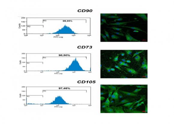
Human mesenchymal stem cells were isolated from adipose tissue with collagenase type I digestion in Dulbecco's modified eagle medium. When ASCs reached a confluence of 80%, they were analyzed using flow cytometry and confocal microscopy in order to confirm the expression of specific stem cells markers CD73, CD 90 and CD105. Analysis was positive for the expression of all markers. The histograms of positive cell markers and confocal microscopy results are given in Figure . 1. All markers were positive in more than 95% and an intense fluorescence in confocal microscopy was also observed.
20. b) NP and AF cells morphology, characterization and
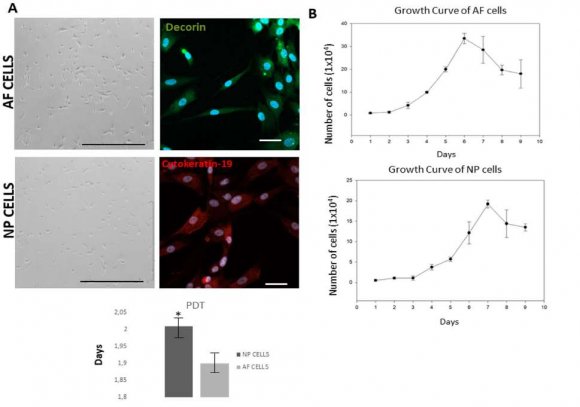
Growth Kinetic NP and AF showed adherent, spindle-shaped fibroblast-like cells (Fig. 2A). AF cells reached 80-90% confluency by days 4-6 after culture, though NP cells were confluent within 9 days after seeding. A confocal characterization of NP and AF cells was carried out. NP and AF cells were markedly positive to cytokeratin-19 and decorin respectively (Fig. 2A).
The growth curve for NP and AF cell populations was characterized by an initial lag phase of 48 hours in AF cells and 72 hours in NP cells followed by a log phase, whereas AF cells immediately grew logarithmically until day 6. NP cells reached this maximum at day 7-8 (Fig. 2B). In order to determine the population doubling time of cells was calculated at passages P1, P3 and P5 (Fig. 2B). Our results showed that the population doubling-time of AF cells increased every 1.90 days and 2.0 in NP cells. We confirmed that the proliferation potential was higher in AF cells than in NP cells. The nucleus was stained with DAPI (blue).
21. c) ASCs derived-CM was able to trigger an immunomodulatory response on NP and AF cells
So as to analyze the immunomodulatory effect of CM on AF and NP cells, inflammation was induced by adding TNF to the culture medium. The concentration of TNF and the incubation time for the optimal response of the in vitro inflammation model were determined by previous experiments in our laboratory. Different concentrations and action times were tested, establishing a TNF concentration of 10 ?g/mL and hours of incubation as parameters with an optimal inflammatory 12 response.
AF, NP and ASCs cells were seeded both with and without TNF/CM and incubated for 12 hours as previously described. RNA extraction and qPCR were then developed to analyze the relative expression of different genes (TNF, IL1-?, IL1-?, IL-6, IL-8 and IFN-?) related to inflammation and therefore discogenic pain. These genes were analyzed under the different conditions to determine the TNF and CM action in this pathology.
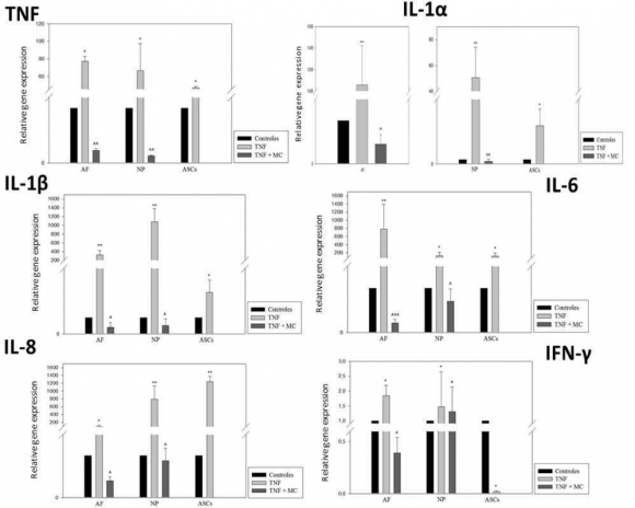
The increase in TNF expression in AF and NP cells when they were stimulated with TNF compared to non-stimulated cells (controls) (*p ?0.05) is given in Figure 4. A decrease was also observed in this expression when we cultured the TNF-stimulated cells with CM (^^p ? 0.01) in both cell types. Surprisingly, an elevated expression of TNF in ASCs where inflammation has been induced was observed and was statistically significant regarding unstimulated ASCs (*p ? 0.05), although the values were lower than in AF and NP cells.
We observed a statistically significant high expression (***p ? 0 .005) in TNF-3 stimulated AF cells with regard to the control of the IL-1? gene as well as a decrease in this expression when the inflamed cells were cultured with MC, which were also statistically significant (^^p ? 0.01) (Fig. 3). A similar pattern was shown in NP cells, observing an elevated expression of IL-1? in ASCs, where the inflammation has been induced, though to a lesser extent than AF and NP cells stimulated with TNF. The difference in stimulated ASCs was statistically significant with regard to non-stimulated ASCs (*p ? 0.05) (Fig. 3).
When IL-1? expression was analyzed, results showed a statistically significant (**p ? 0.01) elevated expression in AF and NP cells stimulated with TNF compared to non-stimulated cells with outstanding NP cells with very high expression. We also observed a decrease in this expression when TNF-stimulated cells were cultured with CM (^p ?0.05) in both cell types. With regard to ASCs, a high expression of IL-1? when inflammation had been induced was observed, and it was statistically significant regarding non-stimulated ASCs (*p ? 0.05), these values being lower than in AF and NP cells stimulated with TNF (Fig. 3).
On analyzing IL-6 and IL-8 results, we observed an elevated expression in AF and NP cells stimulated with TNF with respect to the control (** p ? 0.01). When CM was added, IL-6 and IL-8 expression decreased significantly (^^ p? 0.001). IL-6 expression in TNFstimulated NP cells is also high (* p ? 0.05), but to a lesser extent than in AF cells. With regard to IL-8 The same tendency was shown in both types of cells, though higher values were obtained in NP cells. The same effect was observed in ASCs with TNF, higher values were obtained, which were statistically significant (*p ? 0.05) compared to the non-stimulated ASCs (Fig. 3).
When IFN-? was studied in AF and NP cells stimulated with TNF a considerable increase with respect to the non-stimulated cells was shown. Once again, IFN-? levels were reduced significantly when CM was added in both cell types. Conversely, a low expression, almost nil, of IFN-? was observed in stimulated-ASCs (Fig. 3).
22. d) ASCs derived-CM down regulate neurotrophic factors in NP and AF cells
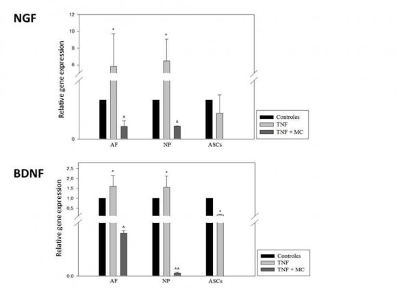
With regard to the neurotrophic factors analyzed, the elevated expression of NGF in AF and NP cells stimulated with TNF with respect to non-stimulated cells (*p ? 0.05) is shown in Figure 4. Thus, we observed a decrease in this expression when CM was added in both cell types. The expression of NGF in ASCs was high in the control group and values were very close to those in the inflamed group.
Finally, analyzing BDNF, cell behavior was equivalent to that observed in response to the other factors. However, the decrease in NP stimulated cells cultured with CM was significantly higher (Fig. 4).
These results are given in Table 2. This table shows the genes analyzed in the in vitro model of discogenic pain, how their expression has developed when AF and NP cells have been stimulated with TNF and how CM is capable of modulating the response.
23. IV. Discusion
Low back pain has become a principal leading cause of work disability in recent decades owing to its high incidence. However, existing treatments are not carried out in a simple enough way. There is therefore an urgent need to establish an effective and simple remedy to treat this disease in the clinic practice [17]. Stem cells have emerged as a key element of regenerative medicine therapies, thus providing numerous potential cell therapies for treating a range of degenerative diseases and traumatic injuries. A recent paradigm shift has come about suggesting that the beneficial effects of stem cells may not be limited to cell restoration alone but may also be due to their transient paracrine actions. Stem cells are capable of secreting potent combinations of trophic factors which modulate the environment of molecular composition to evoke responses from resident cells [18].
In this study, human ASCs were obtained from the infrapatellar fat of patients. Immunofluorescence characterization and culture behavior of ASCs confirmed the characteristics of MSCs, with the expression of all markers being very prominent [19]. These results agree with other authors who had previously characterized ASCs for their easy collection and isolation and for their applications in humans [20]. Furthermore, some authors suggested that ASCs could be a more suitable cell type than bone marrow mesenchymal stem cells for IVD regeneration [21]. The AF and NP cells culture posed no difficulty since they were obtained from stable cell lines and their specific cell markers and growth kinetics were tested. As have been published our results confirmed a higher PDT in AF cells than in NP cells [22]. Confocal microscopy analysis was positive for specific markers. Decorin fluorescence was higher in AF cells and cytokeratin-19 in NP cells [23].
24. a) Immunomodulatory effects of MC on TNF-stimulated AF and NP cells
Discogenic pain entails multifactorial changes occurring with late IVD degeneration which interact with the peripheral nervous system and the central nervous system which cause the pain. Pain may be the result of biomechanical instability, damage to the endplate, inflammation of the nerves or sensitization [24]. Not all degenerated IVDs reveals discogenic pain. However, IVD degeneration is undoubtedly one of the most important keys [25]. In general, there are a vascular response and cell recruitment and activation caused by various cell types. A variety of cytokines have been found in human IVD in varying amounts, depending on whether the IVD is healthy, degenerated or herniated such as TNF, IL-1?, IL-6, IL8, MMPs, among others [26,27].
The aim of this work is to try and detain an irreversible inflammatory cascade at somepoint using ASCs derived-conditioned medium as a therapeutic strategy.
Since TNF is an important pro-inflammatory cytokine in the discogenic pathology, inflammation was induced in all our culture conditions with TNF, although other authors showed equivalent inflammatory effects with INF-? and LPS [28]. When we analyzed the expression of pro-inflammatory genes, we observed a high expression of all of them under inflammatory environment conditions produced by TNF, as we know that it is a key mediator in disc degeneration and low back pain [6]. TNF and both isoforms of IL-1 (IL-1? and IL-1?) showed high expression when stimulating IVD cells with TNF, decreasing this expression when the CM was added to the cultures. These results were similar to those obtained in the study developed by Bertolo and collaborators in which they state that MSCs can modulate the inflammatory state of IVD [29].
IL-6 is one of the cytokines responsible for causing pain in osteoarticular disease and is a major inducer of inflammation together with IL-8 [26,30]. In our study, we observed that in TNF-stimulated ASCs, the expression of these two cytokines is not as low as would be expected because of their immunomodulatory effect. In the case of IL-6 this may be because, although it has been shown to be one of the main interleukins which induce inflammation, its role is currently being debated due to evidence that it may have an antiinflammatory role [31].
With regard to IL-8, we know that it could be associated with the development of root pain and be activated in acute or subacute herniated discs. Constitutively, MSCs secrete hundreds of factors, among them IL-8 with pro-angiogenic effect [32]. Therefore, as an in vitro model of acute inflammation the immunomodulatory capacity of the CM produced by ASCs has been observed in AF and NP cells [33].
IFN-?, an important macrophage activator and an inducer of the expression of Class II major histocompatibility complex (MHC) molecule, is a cytokine which is critical in both innate and adaptive immunity in humans [34]. Its expression increased when IVD cells were cultured with TNF and decreased when CM was added to culture medium. Kim et al. conclude that IFN-? levels are significantly reduced when activated T lymphocytes are co-cultured with MSCs, which indicates an immunomodulatory effect of these cells and, consequently, of the CM tested in our studies [35].
Previous studies have shown that, with degeneration, there is also an upregulation of vascular endothelial growth factor (VEGF) and NGF that can promote neurovascular in growth inside the IVD [36,37]. While the healthy IVD is avascular and aneural, degeneration is thought to induce structural and biochemical changes that contribute to angiogenesis and subsequent disk innervation, which effectively sensitize IVD and resulting in low back pain [3]. Among the molecules presumably involved in hyper-innervation of IVD are growth factors which are members of the neurotrophin (NT) family and are known to be neurotrophic factors. In addition to their role during the development of the nervous system, NTs also play an important role in inflammatory responses and pain transmission [38]. In fact, NTs, in particular NGF, is a peripheral pain mediator, particularly in inflammatory pain conditions. Furthermore, in the sensory innervation of IVD, it has been shown that under normal conditions NGF regulates the expression in nociceptors of a second neurotrophin, a brain-derived neurotrophic factor (BDNF). BDNF is released when nociceptors are switched on, and it acts as a central pain modulator [39]. NGF is regulated under a wide range of inflammatory conditions, and NGF neutralizing molecules should be effective analgesics in many models of persistent pain. Our results confirmed the high expression of these NTs in TNF-stimulated IVD cells, and how CM was capable of decreasing this expression so CM could have the capacity of being a powerful therapy in the research of discogenic pain treatment.
The immunomodulatory role of MSCs is widely described, but taking into account the CM obtained from ASCs studies are scarce [7,40]. Our data showed the immunomodulatory role of ASCs-derived CM reducing inflammation and these effects could be a key element in inhibiting the activity of the molecular factors involved in the process of tissue degradation and possibly pain control in IVD degeneration pathology.
25. Declarations
Ethics approval and consent to participate Not aplicable. Year 2021
26. Global Journal of