1. Botanical Standardization of the Embeli Ribes
Burmf & Possibilities of Species Substitute Syed Asadulla ? , Ramandang ? & Rajasekharan ? EmbeliaribesBURM. F. is an important drug of Ayurveda. Which is considered as multi remedies *with wide* group of Active consistents.Isolated from the berries.
Because of High Commerce, Traders, are subjected to 26 species of substitution, a detailed botanical investigation with macro & microscopical comparison with the drug used under the name of VIDANGA.
Therefore the present study is an attempt to establish macro & Microscopic characteristic of E.R. as well as to Distinguish the species in Chart. Great emphasis is laid on the most diagnostic characters by which each parts of the plant was identified particularly with the macro biological group to which is belongs as in Table no.1, of fruit collection and genuine and substitution (Table no. 2) which gives the value in distinguishing features between species of fruits(berries). As the leaf is first cleared in the solution of Chloral hydrate & lignification was established by the reaction with solution of phloroglucinol followed by a concentrated Hydrochloric acid (C-HCL) to detect the presence of lignin &also mounted for powder microscopy for fruit in dry condition.
The respective photographs were taken with nokiacamera and measurements were taken with camera lucida support and recorded.
IV.
2. Anatomy
Stem -TS of fresh stem shows a circular outline, with a single layer of epidermis covered with a thin cuticle, numerous lenticels, Below the epidermis 2-5 rows of collenchymatous cortical tissue is present and rest of the cortex is parenchymatous containing numerous simple and occasionally oleoresin cells along with compound starch grains & patches of lignified fibers present at fairly regular intervals towards inner cortex, the vascular bundles, cambium, uniseriate medullary rays and pith is seen.
Petiole -TS of petiole is nearly circular in outline with a depression on the adaxial side, a thick wall epidermis, several well developed layers of collenchymas are present beneath the upper epidermis and a sheath of ground tissue, but only2 to 3 layers on Abaxial side.
Ground tissue is parenchymatous, vascular tissue forms an arc that has widely spaced bundles, a few small prismatic crystals of calcium oxalate are present in the ground tissue. Detection of various anatomical features such as tracheids, trichromes, fibers, glands, cork, stomata, pollen grains etc provides important identification clues in leaf & fruits. ER and many of its species are recorded for their botanical characters in Table no.1 and 2.
EmbeliaribesBURM. F. botanical features deals with the pharmacognostical study of leaf and fruits.
VI.
3. Discussions
EmbeliaribesBURM.F. plant character is identified in FRLHT,Bangalore, and soil samples is submitted in GKVK,Soil sciences ,, Bangalore. The results are awaiting and the Tissue culture study is carried out in I.I.H.R. Bangalore results are awaiting.
4. VII.
5. Conclusion
The present statement is to predict the leaf & fruit characters of EmbeliaribesBURM. F.
Thus it is concluded that the above statement can be validated and authenticated on the basis of their macro µscopical characters are the possibilities of substitutes.
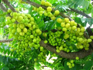
![. Fresh aerial twigs possessing flowers and fruits was collected from I.I.H.R. Hessarghatta fields, Bangalore with accession No.1001. which is an Altitute of 800 meters (2800Sqft) latitude : 13.1323 North, longitude: 77.49332 East. Wind at East 12KM/hr, Temperature (11 0 -25 0 C) Humidity at 45% which is washed thoroughly in running water and the samples were deposited in institutes respository vide voucher specimen numbers 1 to 5 of sample no. 1 (FRLHT Collection No. 55181), dated January 03, 2012, to study and identified the species by Dr. Ravikumar.K. Asst. Director, RMR Division, at Institute of Ayurveda &Integreative Medicine [IAIM], an initiative of FRLHT Herbarium division.andsome of the fresh material is preserved in FAA (Formalin-Acetic acid-Alcohol) and the rest was dried at room temperature and prepared Herbarium preserved, at Research Centre & rest was dried at room temperature for Histological studies,Berry Morphology at Fig.No.1. a) The Drug indicates identification & authentication](https://medicalresearchjournal.org/index.php/GJMR/article/download/422/version/100210/5-Botanical-Standardization_html/3483/image-2.png)
| 9. Sclereid layer of dark Brown of 2-3layers which is | ||||
| tangentially elongated layer thickness ranges from | ||||
| 97.09 µ to 166.44 µ. | ||||
| 10. Epidermis of 5 to 12 layers thick walled narrow and | ||||
| axially elongated cells layer thickness ranges from | ||||
| 41.61 µ to 61.35 µ. | ||||
| 11. In fresh condition Arilluslayer thickness ranges from | ||||
| 485.45 µ to 762.85 µ which is the layer of Testa is | ||||
| covered layer on the external side by thin | ||||
| transparent | rectangular | tangently | arranged | |
| colourless cells or with collapsed parenchyma also | ||||
| 013 | called as membranous Arillus, In dry condition the Arillus is modified into Testa layer modified into | |||
| 2 Year | rectangular fruit(Berries)which scales consist 24 mesocarp and endocarp on powder analysis the in number per on T.S.ofepicarp, | |||
| testa region shows a group of oleoresin cells and | ||||
| stone cells. | ||||
| ( ) B | ||||
| Serial | Regions | Cortex Colour | Testa Fracture | Longitudinal | Special Features |
| No. | Stiations or Scaly | ||||
| markings | |||||
| 01 | Hubli | Ash Brown to green | Gradually Testais Erupted and | 22 | Intermittent eruption |
| Hebsur | few lines are seen. | in 30% of seeds | |||
| 02 | AyurHubli. | Brownish-Black with | Testa is firmly attached | 28 | Few scaly eruption |
| white patches | seen | ||||
| 03 | Himalaya | Brown Green | Calyx, Broken, in 30% | 24 | Not to be seen |
| 04 | Kerala | Brownish Black | 1% of Hemisphere Testa Breaks & | 22 | Nil |
| 99% is safe | |||||
| 05 | Rajastan | Brownish red to Green | Testa is erupted in 50% of seeds | 22 | 4-5 |
| 06 | Fathepur | Matte Ash -Brown | In half Hemisphere | Nil | Single Fracture |
| 07 | FRHLT | Reddish Brown | 50% Testa is broken to powder | 28 scales are found | Scaly depression |
| 08 | Hessarghatta | Reddish Brown to | Testa is attached to the seed | 24 scales are found | Scaly depression |
| Green | |||||
| 09 | AmrutKesari | Brownish Black | Testa is erupted to 25% | 24 | 2 to 3 |
| Sl. No. | Drug Name | Authour Name | Source | Uses | Published | ||||
| 01 | EmbeliaribesBurm.f. | Chua,LSL; | JLCHForest Research | Anthelmintic | Plant resources of South-East Asia No. 12(1): | ||||
| van Valkenburg, | Institute | Malaysia, | Medicinal and poisonous plants 1; de Padua, | ||||||
| Jalan FRI, Kepong, | L.S., Bunyaprapatsara, N &Lemmens, R.H.M.J. | ||||||||
| 52109 Kuala Lumpur, | (eds); Paperback edition; Bogor, PROSEA | ||||||||
| Malaysia | Foundation, 1999; p 257-258 | ||||||||
| 01a | EmbeliaribuBurm.f. | Chua ngutVo Van Chi | Vietnam(grows | in | ripped fruits Treat | Vietnamese Medicinal Plants], Hanoi, Medicinal | |||
| Tudien cay thuoc | waste | land, | hill | bitten by snack, earth | Publ. House, 1997; p. 244. | ||||
| mountains) | worm, whites, cough | ||||||||
| and diarrhoea. | |||||||||
| 02 | EmbeliarobustaRoxb.(Vir | Chua, | LSL; | van | |||||
| anga. Birang-i-kabuli,) | Valkenburg, | ||||||||