1.
Shoulder and Elbow joint, its cutaneous supply includes skin over anterolateral aspect of fore arm till the base of Thenar eminence. [1] II.
2. Materials and Methods
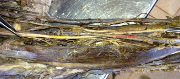
Forty limbs(Rt: 20; Lt: 20) from 20 embalmed cadavers were utilized during the study period of three years. The pectoral region,the axilla, the arm, cordsand the branchesofthe infraclavicular part of the brachial plexuswere dissected. The variations of median and musculocutaneous nervewere noted.
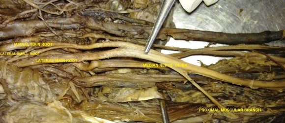
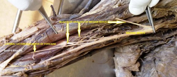
Observations: The musculocutaneous nerve was absent in 2 upper limbs of the same cadaver, median nerve showed varied anatomical pattern in respect to origin, course and termination. In left upper limb, the musculocutaneous nerve was found absent and median nerve was arising from three roots (2 from lateral cord and 1 from medial cord of brachial plexus), during its course in arm it was observed that nerve giving one proximal and one distal branch. Proximal branch supplying coracobrachialis, biceps brachii, and brachialis. Distal branch arise from lateral side of nerve and passes below the biceps brachii to continue as lateral cutaneous nerve of forearm.
3. Discussion
Multiple variations of Brachial plexus havebeen documented by Henry Hollinshead in 1969 [2] . Uzan [3] found three Roots from Lateral Cord and one Root from Medial Cord. These roots united to form Median nerve.
In the present study one specimen shows two roots from lateral cord and one from medial cord. Jahanshahi M [4] described absence of Musculocutaneousnerve and muscles normally supplied by it were supplied by Median nerve, however the Median nerve was formed in normal way. In our case Median nerve has three Roots, which is a variation, as Median nerve is normally formed by two Roots.Satyanarayan N [5] describes three unilateralcases of variations in the formation of Mediannerve. In the first case, the Median nerve was formed on the medial side of Axillary artery and also at a higher level. Later the Median nerve continued behind the Brachial artery and received a communicating branch from Lateral Cord of Brachial plexus. In the second case, formation of Median nerve was by three Roots, two Roots from Lateral Cord and one Root from Medial Cord. In the third case, Median nerve was formed by four Roots, three Roots from Lateral Cord and one root from Medial Cord. We have found three Roots of Median nerve in one left limb specimen which was in agreement with second case of above mentioned author. The musculocutaneous nerve (C4-C6), a mixed peripheral nerve, arising from the lateral cord of the brachial plexus in the axilla, usually innervates the muscles of the anterior compartment of the arm and then continues as the lateral cutaneous nerve of the forearm [6] . PrasadaRao [7] reported two cases of absent musculocutaneous nerve from the lateral cord of the brachial plexus. In the present study, the absence of the musculocutaneous nerve was observed in 2 specimens of same cadaver. Ihunwo et al [8] reported a case of the bilateral absence of the musculocutaneous nerve from the lateral cord of the brachial plexus, with four branches arising from the lateral sideof the median nerve. This report was in correspondence with that of the present study with little anatomical variations.Combination of absence of Musculocutaneousnerve and three roots of Median nerve as seen in the present case is a rare occurrence. Knowledge of this variation is crucial while performing block dissection of Axilla, reconstructive flap surgeries, treating Humeral fractures by open reduction and even while performing incision and drainage of an Axillary abscess. Presence of such variation should always be kept in mind while testing of muscle after administration of neuromuscular block.
4. a) Embryological Explanation
William Larsen [9] quotes that ventral column motor axons sprout from spinal cord in craniocaudal direction around day 30 in a developingembryo. An apical structure "Growth Cone" is formed at the growing tip of axon. The Growth Cone decides the path to reach the target organ. Filopodia present on Growth Cone grow towards the target organ by sensing molecular markers secreted by surrounding tissue. Location and innervations of the target organ (muscle, joint, skin) is dependent on secretion of certain tropic substances by target organs and its identification by growing axon. Absence of Musculocutaneous nerve in the present case can be explained that growth cone Filopodia of ventral column motor axon sprouting from C5, C6 and C7 spinal segments took an unusual path and travelled from Lateral Cord to form Median nerve via Lateral Root of Median nerve. However the growth cone recognized Median nerve had two medial roots because fibres from C8 and T1 spinal segments joint lateral root of median nerve separately.
IV.
5. Conclusion
Roots of brachial plexus seen in root of neck in way to axilla where they form chords. So any surgeries involving axilla and posterior triangle of neck needs utmost care and sufficient knowledge of formation of median nerve and variation in origin of musculocutaneous nerve. Although musculocutaneous nerve found absent in bilateral side of specimen surgeon should need to know about multiple variation of this nerve which have clinical significance in posttraumatic evaluations and exploratory innervations of the arm for peripheral nerve repair. Even though median nerve is main nerve of forearm it supplies arm when absence of musculocutaneous nerve, so it is necessary to gain awareness about such variations before any interventions in treatment of fracture of Humerus and surgeries related to elbow joint.