1. Introduction
he conventional method of diagnosing ascariasis is by testing the stool for the presence of the eggs. When there are atypical abdominal symptoms a routine ultrasound scan using the common 3-3.5 MHz probe yields no definite findings to diagnose intestinal ascariasis. If a high frequency probe of 5-10 MHz is used instead, intestinal ascariasis could be definitely established. This case illustrates a typical worm diagnosed with the help of a high frequency probe.
2. II.
3. Case Report
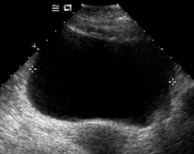
A 17-year-old boy was referred from emergency for ultrasound scanning of abdomen because of vomiting and generalized abdominal pain mainly in the periumbical region of short duration.There was no history of fever and no sign of peritonitis. The routine abdominal scanning using a 3-3.5 MHz probe was inconclusive. Another attempt using a high-density multifrequency linear probe of 5-10 MHz showed an entirely striking picture. There was brighter tubular shadow having an average diameter of 4 mm with a central hypoechoic core (Fig. 1). A cross-sectional view of the worm typical bright ring shadow(Fig 2).. These structural features were consistent with the diagnosis of intestinal ascariasis. The other visceral echoes were normal.
4. III.
5. Discussion
Ascaris lumbricoides is a common nematode infesting a major percentage of human beings worldwide (more than 1.4 billion). It grows to a maximum length of 35 cm. This species is host specific to human beings and lives longer (1-2 years) with in the small intestine. Infested individuals are mostly asymptomatic though it is a causative agent for some very common symptoms. The literature shows enough reports on biliary ascariasis 1,2,3,4 .The ultrasound scanning is the specific diagnostic tool in case of biliary infestation. Intestinal ascariasis demands the use of a higher frequency high-density probe of 5 -10 MHz, as illustrated here. The live worm on longitudinal section appears as a writhing tubular shadow having brighter margins described by some as 'strip sign'. There is a hypoechoic core producing the 'inner tube sign'. The cross-sectional picture is also characteristic of a tubular body described as the ring sign or bull's eye sign if seen with in the CBD or a narrow lumen 2 . when the crowded worms form a ball like mass, the ultrasound sectional view can be called as the "stacked tubes sign" 5 .
Fig. 1 : A longitudinal section of segment of Ascaris lumbricoides shows the tubular shadow with brighter parallel walls (strip sign). The core is hypoechoic(inner tube sign). The patient's bowel walls are also identifiable