1. I. Introduction
s the blast injuries are deliberately devised to inflict maximum damage, these injuries often occur in crowded places like markets and the range of people effected varies greatly as regards age, sex, number of people injured .We present a case of an young individual who received more than 75 pellet injuries that were scattered throughout the body. Most of the pellets were received on the right upper limb with a few in chest, abdomen and pelvis. The neck fortunately had pellets in safe areas .This was one rare case of trauma in which so many pellets were received by a single patient. In this patient the vitals were Normal. Neurological examination was normal as was Orthopaedic examination with evidence of bleeding from multiple sites on the body which was taken care of during the initial assessment. However there was no serious source of bleeding. Trauma in Kashmir valley has taken a toll. Many people have died, some have got lifelong disabilities and some are incapacitated. Neck: The area of the neck is especially Important as this area is unprotected by bone or dense muscular covering. The most significant neck injuries result from penetrating trauma, blunt neck trauma does occur and can be particularly difficult to manage because it often involves the airway, the first priority in trauma care. Fatality rates for penetrating neck trauma range up to 50% for rifle or shotgun blasts.
The neck is divided into a number of anatomic triangles. Two large triangles are important in discussing penetrating neck trauma. Penetrating wounds that enter through the sternocleidomastoid muscle or anterior triangle carry a high likelihood of significant vascular, airway, or esophageal injury. In contrast, wounds to the posterior triangle rarely involve the esophagus, airway, or major vascular structures, but, if directed inferiorly, intrathoracic injury can occur.
The boundaries of Anterior triangle of neck Anterior: anterior median line of neck Posterior: anterior border of sternomastoid Base: base of mandible and a line joining angle of mandible to mastoid process.
Apex: manubrium sterni. Zone III : It is the part of the neck above the angle of the mandible. The risk of injury to the distal carotid artery, salivary glands, and pharynx is greatest in this zone, and exposure can be particularly difficult.
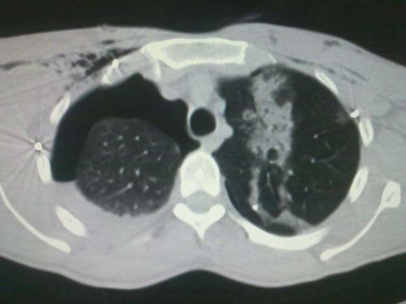
2. III. Thorax
The life threatening injuries incurred in penetrating trauma are distinctly different from those of blunt injuries. Penetrating thoracic injuries (e.g., stab wounds, gunshot wounds, and impalement on a foreign body) primarily injure the peripheral lung, producing both a hemothorax and pneumothorax. More than 80% of all penetrating chest wounds cause a hemothorax, and nearly all cause a pneumothorax. The Penetrating injuries that enter or traverse the mediastinum must also be evaluated for potential cardiac, great vessel, or esophageal injury. Hemodynamically unstable patients with mediastinal entering or traversing wounds should be considered to have exsanguinating thoracic hemorrhage, pericardial tamponade, or tension pneumothorax.
Hypovolemia from intrathoracic hemorrhage is second only to rib fractures as a sequel of thoracic trauma. Penetrating injuries resulting in direct cardiac injury and pericardial tamponade can rapidly compromise cardiac function.
3. IV. The Abdomen
Is the third most commonly injured body region. Abdominal injuries can be particularly challenging, because it is often difficult to assess intra-abdominal pathology in the multiple-injured victim. Blunt trauma continues to be the most common mechanism of injury to the abdomen. Abdomen is divided into nine regions by: ? Two vertical planes: right and left lateral planes.
Passing from midinguinal point and crossing tip of ninth costal cartilage (mid-clavicular lines) Stomach: It lies obliquely in upper and left part of abdomen, occupying epigastric, umbilical and left hypochondriac region. Most full-thickness gastric injury is due to penetrating trauma. Gastric rupture secondary to blunt trauma is rare . If vomitus or gastric aspirate is bloody, an injury to the stomach should be suspected. However, it is not unusual for small amounts of blood to be found in the gastric aspirate, even though laparotomy reveals no grossly apparent lesion.
Duodenum: The position of duodenum:
Duodenum lies above the level of umbilicus against L1-3 vertebrae, extending ½ inch to right and 01 inch to left of median plane. The length of duodenum and its different parts Duodenum is a 10 inch long, curved around the head of the pancreas in form of C. Approximately three fourths of duodenal injuries result from penetrating trauma, and one fourth are from blunt injuries. Penetrating injuries are usually readily diagnosed at operation, but the insidious nature of many blunt duodenal injuries makes the initial diagnosis difficult.
In addition to maintaining a high index of suspicion in patients with appropriate injury mechanism, a serum amylase level should be initially obtained and if elevated, repeated at 6-hour intervals in patients with blunt abdominal trauma.
The radiologic signs of duodenal injury on the initial plain abdominal or upright chest radiograph are subtle, showing only mild scoliosis, obliteration of the right psoas muscle, or retroperitoneal air that is difficult to distinguish from the overlying transverse colon.
Pancreas: Pancreatic trauma is relatively uncommon, accounting for less than 10% of all abdominal injuries. Although the pancreas is relatively protected in the retroperitoneum, the increasing frequency of large-caliber, high-velocity civilian gunshot wounds contribute to an increasing incidence of pancreatic injury. Because the pancreas is surrounded by major abdominal organs and blood vessels, associated injuries are common. Liver and biliary tree: Liver. The liver is the most commonly injured organ following penetrating trauma and the second most commonly injured organ following blunt trauma. Fortunately, the injury is often minor and easily managed. When the liver is the only organ injured, half the lacerations are nonbleeding and do not require suture. In most lacerations that are bleeding, the source is within the substance of the liver, and control can be obtained by direct ligation. With deeper lacerations, bleeding may initially be so significant as to prevent adequate exposure. Under these conditions, the next maneuver is that of inflow occlusion (Pringle maneuver).
Porta Hepatis: It is a deep transverse fissure, 05 cm long, on the inferior surface of right lobe of liver, between quadrate lobe below and front and caudate lobe above. Through it vessels, nerves and ducts pass to and from liver Isolated penetrating or blunt injuries to the porta hepatis are uncommon. Unless there are associated injuries to the aorta and inferior vena cava (which take priority) noted at laparotomy, an immediate Pringle maneuver frequently allows isolation of the structures in the porta hepatis and determination of whether the injury involves the portal vein, hepatic arteries, or common bile duct. Repair of vascular injuries takes precedence over biliary structures. Injuries to the portal vein have a high mortality rate. The portal vein supplies 80% of the total hepatic blood flow and 50% of the oxygen delivery.
Gallbladder: injury to this organ has been reported to occur after both penetrating and blunt trauma. Cholecystectomy is the procedure of choice for severe contusion, avulsion, or perforating injuries to the gallbladder.
Spleen: The spleen lies obliquely along the long axis of 10th rib. It lies mainly in left hypochondrium but the posterior end extends into epigastrium. It is directed downwards, forwards and laterally. The spleen is the most commonly injured intraabdominal organ. Splenic injury must therefore be suspected in any patient with blunt abdominal trauma, particularly if associated with left lower rib fractures. The diagnosis is often suspected on physical examination but is generally confirmed by abdominal CT scan or exploratory celiotomy for hemoperitoneum.
Colon: The colon is second only to the small bowel in frequency of abdominal organs injured from gunshot wounds, and third (liver, small bowel) following abdominal stab wounds. Gunshot wounds to the abdomen generally require exploratory laparotomy.
Kidneys: Penetrating injury to the kidney may be secondary to a gunshot. Parenchymal injury caused by a low-velocity weapon is usually not life threatening and is generally easily treated with débridement and primary repair of the kidney with dependent drainage. Highvelocity bullet wounds are different. Many nephrectomies and partial nephrectomies are performed in patients with high-velocity penetrating injury because of inability to control hemorrhage and accurately define the extent of the injury.
4. V. Conclusion
It can be safely concluded that the proper knowledge of Anatomy is important for first recognizing the injuries especially be pellets or blasts. The perfect knowledge of Anatomy is helpful in management as well. As such a surgeon or a trauma orthopedic surgeon should be well aware of the important anatomic relations in the neck, thorax and abdomen.


| Year 2014 | |
| Volume XIV Issue III Version I | A |
| D D D D ) H | |
| ( | |
| The subdivisions of Anterior triangle | |
| Submental | |
| Digastrics | |
| Carotid and | |
| Muscular triangle. | |
| The boundaries of Digastric triangle | |
| Anteroinferiorly: anterior belly of digastrics | |
| Posteroinferiorly: posterior belly of digastrics | |
| stylohyoid. | |
| Base: base of mandible and a line joining angle of | |
| mandible to mastoid process. | |
| Roof : skin | |
| Superficial facia: has platysma and cervical branch of | |
| facial nerve. | |
| Deep fascia: splits to enclose submandibular gland. | |
| Floor: mylohyoid, Hyoglossus and Middle constrictor |