1.
To discuss the condition of a child patient with abdominal pain related to autoamputated appendix.
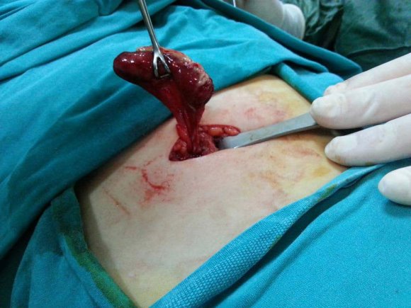
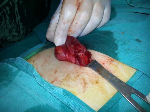
A male patient at 12 has admitted to our clinic with abdominal pain and vomiting complaints lasting for two days. His white blood cell (WBC) count was 9,100 and C-reactive protein (CRP) level was 27,89. Although direct radiographs have reflected normal structure, there was a diffuse thickening on ileum and there was a peripheral fluid collection surrounding the caecal walls. The patient was hospitalized. His abdominal pain has temporarily relieved but, he has undergone surgical intervention as his abdominal pain has exacerbatively recurred at the end of day one. During intraoperative exploration, we have observed that the appendix was totally seperated from caecum and its mesenteric perfusion was partially deteriorated (Fig. 1). Appendectomy operation was performed without caecal suturation (Fig. 2) and our patient was discharged from hospital two days after the operation with total remission.
2. GJMR-I Classification: NLMC Code: WI 535
3. AutoamputatedAppendixACaseReport
Strictly as per the compliance and regulations of:
b) Case Report Introduction a) AimTo discuss the condition of a child patient with abdominal pain related to autoamputated appendix.
4. b) Case Report
A male patient at 12 has admitted to our clinic with abdominal pain and vomiting complaints lasting for two days. His white blood cell (WBC) count was 9,100 and C-reactive protein (CRP) level was 27,89. Although direct radiographs have reflected normal structure, there was a diffuse thickening on ileum and there was a peripheral fluid collection surrounding the caecal walls. The patient was hospitalized. His abdominal pain has temporarily relieved but, he has undergone surgical intervention as his abdominal pain has exacerbatively recurred at the end of day one. During intraoperative exploration, we have observed that the appendix was totally seperated from caecum and its mesenteric perfusion was partially deteriorated (Fig. 1). Appendectomy operation was performed without caecal suturation (Fig. 2) and our patient was discharged from hospital two days after the operation with total remission.
5. Autoamputated Appendix -
6. Discussion
Appendix is derived from midgut and first appears during the 8 th week of the embryonic development as an outpouching of the cecum and consequentally rotates to medial position with gut rotation. 1 The mean length of the appendix is between 8-10 cm. Congenital absent appendix is a very rare condition so that even many very experienced surgeons have not encountered. Pester has reported few cases with absent appendix. 2 If the lower cecal segment do not undergo a thinning process, this may lead to total hypoplasia or absence of the appendix. 3 There are only a few cases in the literature reporting absence of appendix. 4 It is considered that the absence of appendix is associated with autoamputation. However, there is no such case which exhibits appendix autoamputation during surgical intervention so far. Thus, the current case represents the first autoamputated appendix case during surgical intervention. It may be strongly possible that perfusional disorders around the autoamputated appendix could give rise to a necrotic seperation of the appendix from cecum by time, because we have observed that appendicial tissue has an ischemic appearance with changed color to dark purple/black.
7. III.
8. Conclusion
It is noteworthy that appendix autoamputation was intraoperatively observed in this case. Our case may contribute to more clearly reveal the underlying ethiopathogenesis in absent appendix.