1. Introduction
pine provides structure for human body. It undergoes a process of changes with age, stress and strains which causes various spinal problems like disc degeneration, disc herniation, vertebral compression fractures and bone spurs. The IVDs are the joints of the spine having load bearing structure [1]. They lie between the vertebrae bodies and are separated from them by a hyaline cartilage endplate. The IVD consists of the inner ring called Nucleus Pulpous (NP) surrounded by an outer ring called Annulus Fibrosis (AF). These structures differ in functional and molecular properties [2]. Nucleus pulpous has high water content and is compressible. It is essentially a semi-fluid like structure, where water and proteins make it, almost, in compressible substance. Reduction in the water content from NP leads to a loss of IVD height. Exposure to heavy mechanical stress over long periods is thought to be a factor leading to Inter Vertebrae Disc Degeneration (IVDD) [3]. The IVD does not possess self-repair capacity. Degeneration affects all areas of IVD, but some evidence indicates that the most noticeable changes occur in the NP.IVDD problem is the most serious problem because lower back pain is strongly associated with it [4][5][6]. IVDD is treated as serious because most of the patients are suffering from this problem [7]. Disc degeneration also trigger other problems like neck pain, numbness, tingling, loss of muscle strength, walking and standing difficulty and paralysis [8].
Backbone anatomical structure detection and labeling is a necessary step for various analysis tasks of the vertebral column such as IVDD, disc herniation, disc bulging.
Appearance, shape and geometry measurements are necessary for abnormality detection locally at each disc and vertebrae as well as globally for the whole spine [9]. Manual step in detecting IVDD can be time consuming and will depend on the experience of the Radiologist/Doctor. Minor IVDD problems are difficult to detect manually. In some cases, if manual intervention is restricted to picking a set of points, errors are unlikely to occur and it does not take much time. However, it is desirable to automate the detection of IVDD and avoid the manual intervention especially if the size of the workload is large.
Automation of IVDD diagnosis reduces the large burden on radiologists who have to diagnose hundreds of cases each day using clinical MRI. Segmentation of MR images is a complicated task as there is no unique correspondence of grey-level ranges to different tissue types [10,11].
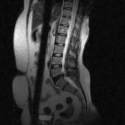
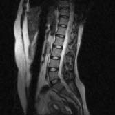
The segmentation of IVD is a prerequisite for the Computer Aided Diagnosis (CAD) of disc degeneration, while it could also be useful for computer based planning prior to spinal surgery [12]. In T2-weighted sagittal MR images a normal disc appears like a bright ellipse because it contain more water content surrounded by a dark ring, whereas a degenerated disc appears darker and often has an irregular shape. These case are shown in fig 1. These features are used in Machine Learning techniques that have been widely and successfully used in CAD [13][14][15]. A novel method is proposed in this paper to detect disc degeneration, disc compression. In proposed method, the intensity values of the inter vertebrae, length width values are extracted to create a template. VESTAL [16] extracts the spinal canal from MR image it guides the proposed method to detect the vertebrae and IVD to extract IVD features.Normal image spinal canal appeared as bright and the path near the vertebrae body also continuous having smooth transition. In abnormal case the problematic disc appeared as dark and the spinal canal near this region also appeared as dark.
2. A) Normal Image b) Abnormal image
Figure 1 II.
3. Related Work
So far, most of the studies that deal with the quantification of disc features for diagnostic purposes have been based on manually segmented [17,18]. In addition, very few researchers report on the automatic segmentation of IVDs. Labeling vertebrae and IVD plays key role to detect the IVDD.A framework has been designed by Tobias et al., that takes an arbitrary CT image, e.g., head-neck, thorax, lumbar, or whole spine, as input and provides a segmentation in form of labelled triangulated vertebra surface models [19]. This framework has been tested on 64 CT images even including pathologies. In 56 cases, it was successfully applied resulting in a final mean point-to-surface segmentation error of 1.12 ± 1.04 mm. One key issue is a reliable identification of vertebrae. For a single vertebra, they achieved an identification success of more than 70%. Increasing the number of available vertebrae leads to an increase in the identification rate reaching 100% if 16 or more vertebrae images.
Samuel et al., introduced a novel approach for segmenting articulated spine shape models from medical images. A nonlinear low-dimensional manifold is created from a training set of mesh models to establish the patterns of global shape variations. Local appearance is captured from neighborhoods in the manifold once the overall representation converges. Inference with respect to the manifold and shape parameters is performed using a higher-order Markov random field (HOMRF). Clinical experiments demonstrated promising results in terms of spine segmentation. Quantitative comparison to expert identification yields an accuracy of 1.6± 0.6 mm for CT imaging and of 2.0± 0.8 mm for MR imaging, based on the localization of anatomical landmarks [20].
Yiebin Kim et al., proposed a fully automatic method for vertebrae segmentation in the CT volume data. The method constructs 3D fences that separate adjacent vertebrae from valley emphasized Gaussian images. Initial curves for the 3D fences are extracted from intervertebral discs, detected with anatomical characteristics, and then optimized using a deformable model [21]. A minimum cost path finding method corrects any erroneous curves trapped into a local minimum. Final volume is labeled with help of the 3D fences by a fence-limited region growing method. This method has been applied to 50-patient data sets and has proved to be very successful.
Szu-Hao Huang et al., developed a fully automatic vertebrae detection and segmentation system. To produce an efficient and effective vertebrae detector, a statistical learning approach based on an improved AdaBoost algorithm is proposed. A robust estimation procedure is applied on the detected vertebra locations to fit a spine curve, thus refining the above vertebra detection results.In their implementation, the proposed AdaBoost-based detector is trained from 22 spinal MR volume images. The experimental results show that the proposed vertebra detection and segmentation system achieved nearly 98% vertebra detection rate and 96% segmentation accuracy on a variety of testing spinal MR images [8].
Kelm et al., proposed a fully automatic and robust approach for an automated scan alignment as well as for the segmentation and analysis of spinal disks and vertebral bodies in Computer Aided Diagnosis applications [22]. Experimental results based on 42 MR and 30 CT volumes show that their system not only achieves superior accuracy but also is among the fastest systems of its kind in the literature.A two-level probabilistic model for the localization of discs from clinical Magnetic Resonance Imaging data that captures both pixel-and object-level features was proposed by Raja et al. They used generalized expectation and maximization for optimization, which achieves efficient convergence of disc labels. Their two-level model allows the assumption of conditional independence at the pixel-level to enhance efficiency while maintaining robustness. A dataset that contains 105 MRI clinical normal and abnormal cases for the lumbar area were used and thoroughly tested their model and achieve encouraging results on normal and abnormal cases [9].
Max et al., proposed an unsupervised intervertebral disc segmentation system based on middle sagittal spine MR scans. This system employs the novel anisotropic oriented flux detection scheme which helps distinguish the discs from the neighboring structures with similar intensity, recognize ambiguous disc boundaries, and handle the shape and intensity variation of the discs [23]. The information is employed in a set of image descriptors, which jointly constitute an energy functional describing the desired disc segmentation result. The energy functional is minimized by a level set based active contour model to perform disc segmentation. This system is evaluated using a database consisting of 455 intervertebral discs extracted from 69 middle sagittal slices.
4. Volume XV Issue 1 Version I Year 2015 ( D )
A unified framework was presented by Claudia Chevrefils et al., for automatic segmentation of intervertebral disks of scoliotic spines from different types of magnetic resonance image sequences. This method exploits a combination of statistical and spectral texture features to discriminate closed regions representing intervertebral disks from background in MR images of the spine. A total of 22 texture features (18 statistical and 4 spectral) are extracted from every closed region obtained from an automatic segmentation procedure based on the watershed approach. This method is validated using a supervised k-nearest neighbor classifier on 505 MR images coming from three different scoliotic patients and three different MR acquisition protocols [24].
A model, to study the cause of degenerative disc disease is diagnosed by Magnetic Resonance Imaging using artificial neural networks method for training and classification is proposed by Unal et al [25]. In this model cropped sample images of size 200x80 pixels regions were used. From grey level formatted MR images features were extracted using the wavelet transform. These obtained feature vectors are sent to multi-layered perceptron artificial neural networks as an input in order to make a classification. As a result of classification process, intervertebral degenerative disc disease is diagnosed with99.79% accuracy in 67 iterations using12 patients' images.
The 2D semi-automatic segmentation of both normal and degenerated lumbar IVDs from T2-weighted mid sagittal MR images of the spine was proposed by Sofia et al [26]. This task is challenged by partial volume effects and overlapping gray-level values between neighboring tissue classes. To overcome these problems three variations of atlas-based segmentation using a probabilistic atlas of IVD were developed and their accuracies were quantitatively evaluated against manually segmented data. They achieved dice similarity indexes of this method were 91.6% for normal and 87.2% for degenerated discs.
Raja et al., presented a method for automatic diagnosis of lumbar disc herniation using appearance and shape features. They jointly use the intensity signal for modeling the appearance of herniated disc and the active shape model for modeling the shape of herniated disc. They utilized a Gibbs distribution for classification of discs using appearance and shape features. They used 33 clinical MRI cases of the lumbar area for training and testing both appearance and shape models. They achieved over 91% accuracy in detection of herniation in a cross-validation experiment with specificity of 91% and sensitivity of 94% [27].
Shijie et al., proposed a framework on analyzing disc shapes based on a geodesic metric in an anatomical shape space. All disc shapes, containing both normal and abnormal ones, are formulated as elements in this space. Their experimental results demonstrationed a reasonable accuracy of classifying normal and abnormal intervertebral discs. They concentrated on the IVD shape study. The normal disc shapes are generally regular and similar to each other while the abnormal ones suffer various non-rigid deformations [28]. Gocmen et al., calculated the concavity index for each lumbar vertebra in adults, as well. Concavity index was established for each vertebral body by dividing the "central" vertebral height by the anterior vertebral height [29].
An accurate and automated method to detect the abnormal disc presented by Ming-chi and Cheng-An Fang. This method uses two standard models in conjunction with the threshold value to accurately identify the cartilage. Ming-chi and Cheng-An Fang, smooth out the images via morphological methods and find out the average height of the cartilage before they judge, if a certain cartilages are lower in height than the normal range. In comparison with the professional physician's manual segmentation, their image segmentation shows high accuracy, with the highest rate reaching 99.88% [30].
Roberts et al. [31] employed watershed techniques for automatically detecting and segmenting non degenerate intervertebral discs from a combination of proton density (PD) and T2-weighted MR images of the lumbar spine. Chevrefilset al. [32] combined watersheds with morphological operations to segment the discs from thoracic spine MR images acquired utilizing the multie-cho data image combination (MEDIC) sequence. Shilet al. [33] utilized the Hough transform to detect the spinal cord from whole spine MR images, and then located and segmented the discs with a selfadaptive window and edge detection methods. Wachteret al. [34] reported on the segmentation of cervical intervertebral discs from both T1-and T2weighted MR images, utilizing active shape models and fuzzy connectedness methods, with promising results.
5. III.
6. Methodology
To classify the degenerated disc, we are extending our work proposed in [16]. The proposed method labels the vertebrae and IVD using VESTAL algorithm. Then this method detects the abnormal region and identifies the severity of IVDD by measuring features of degenerated disc. To detect degenerated disc, proposed method performs the following five major subtasks.
7. a) Spine curve extraction
The MR images are scaled to uniform size. Apply Weiner filter to preprocess the images to remove noise. Canny algorithm is applied on preprocessed images to detect spinal canal path easily. The spinal canal path is detected using Spinal Canal Path Search (SCPS) method. Algorithm : SCPS() //IM []][] vector contain image information //m*n size of the image //img[][] is used for storing path of the spinal canal //p, q intensity range of values of the spinal canal // imgIntensity[][] intensity matrix Comment only the detected region by canny is used for processing to optimize image processing
x = 0, y = 0, img[m][n] = {{0};{0}}; i =IM. getPixel(x,y) if ( p<i and i< q ) img[x][y] = 1; then imgIntensity[x][y]=i; SCPS(x+1,y) SCPS(x,y-1)8. b) Vertebrae Detection
Location of vertebrae can be easily detected using spinal canal. This process saves time and reduces processing complexity. Only vertebrae regions are searched for detecting vertebrae. Algorithm:VD() //IM vector contains image information //m*n size of the image //img[][] is used for storing Vertebrae values //p, q intensity range of values of the Vertebrae // spinal canal is processed in any direction
// imgIntensity[][] intensity matrix img[m][n] = {{0};{0}}; i =IM. getPixel(x,y) if (p<i and i< q) img[x][y] = 1; imgIntensity[x][y]=i; then VD(x+1,y) VD (x,y+1) VD (x-1,y) VD (x,y-1)The vector img[][] contains vertebrae information and other pixels are not having intensities within the range of vertebrae.
9. c) Intervertebral Disc Detection
Form 3.2 the locations of intervertebral discs are detected. Intervertebral discs are in between these vertebrae. The region is detected using IVDS() algorithm which is similar to VD().These regions can be detected from the breaks in the intensities of the spinal canal and vertebrae. The shape of the IVD can alsobe detected using shape of vertebrae.
10. d) Labeling Vertebrae and IVD
Based on statics features extracted using above procedure 3.2 and 3.
11. e) Detecting Degenerated Region and classification of the MR Image
If spine image contains any abnormality, it can be detected using MR images using step 3.4 if image contains abnormal IVDs. These locations are regions to be considered as degenerated regions.The abnormal disc lost its shape and water content and appearance. So intensity values of abnormal disc generally lower than healthy disc. To detect abnormality in disc the above procedure is applied by considering changes in the intensity range for patient image. The calculated value are used for template matching to detect abnormality region. Based on the result image can be classified as normal and abnormal. If the image is abnormal image template matching procedure results specifies the mismatched region. Based on damage and properties of IVD also it classify the type of problem as degenerated or disc thinning. From above results we conclude that the vertebrae and intervertebral widths are in increasing order for lumber vertebrae except for L5. The brightness of the IVD, width and height is increasing order with age from childhood to middle age. The bright ness decreases as age increases from over middle age. The statistical properties vary based on height, nature of the work done by patient.
12. IV.
13. Results
14. MR
V.
15. Conclusions
In this paper, a novel method is proposed to extract feature of IVD to classify the MR images. The proposed method uses statistical properties and using these properties a template is created. The MR images are classified by comparing extracted feature of the image with template. Proposed method produces 94% accuracy in classifying MR images as normal and abnormal and further it classify degenerated or thinning. This method is very useful for detecting degenerated disc. In future this work can be extend for detecting other disc related problems like disc bulging and disc herniation.


| Algorithm:Template() |
| //m*n size of the image |
| //imgAvg[][] intensity average values image |
| // imgIntensity[][] intensity matrix |
| // N number of sample image for specific age group |
| for k= 1 to N |
| begin |
| fori= 1 to m |
| begin |
| for j=1 to n |
| begin |
| imgAvg[i][j]+=imgIntensity[i][j] |
| end |
| end |
| imgAvg[][]/=N |
| end |
| Vector img[][] contains location information |
| about IVDs. |
| The parameters measured for cervical vertebrae, | VESTAL are shown in table 1, 2, 3 and 4 and their |
| cervical IVD, lumber vertebrae and lumber IVD using | corresponding relation in figure 3, 4, 5 and 6. |
| Vertebrae No | Anterior width | Posterior width | Length | Height |
| C2 | 20 | 20 | 28 | 18 |
| C3 | 21 | 21 | 28 | 19 |
| C4 | 22 | 21 | 29 | 20 |
| C5 | 24 | 26 | 29 | 22 |
| C6 | 26 | 26 | 30 | 25 |
| C7 | 28 | 27 | 32 | 25 |
| Vertebrae no | Anterior width | Posterior width | Length | height |
| L1 | 27 | 26 | 36 | 28 |
| L2 | 29 | 28 | 37 | 29 |
| L3 | 29 | 28 | 40 | 30 |
| L4 | 29 | 26 | 41 | 30 |
| L5 | 28 | 26 | 41 | 29 |
| IVD No | Anterior width | Posterior width | Length | Height |
| L1/L2 | 11 | 9 | 40 | 12 |
| L2/L3 | 13 | 10 | 43 | 13 |
| L3/L4 | 14 | 9 | 52 | 14 |
| L4/L5 | 18 | 11 | 33 | 16 |
| L5/S1 | 22 | 11 | 45 | 16 |