1. Introduction
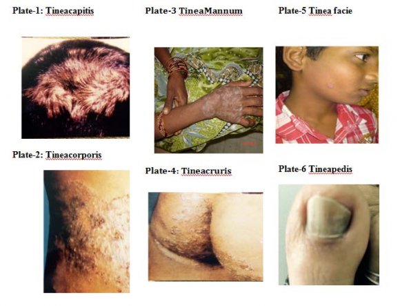
ermatophytoses are a superficial cutaneous mycoses confined to the outer layers of skin, hair, and nails, and do not invade living tissues. The fungi are called dermatophytes. Dermatophytes, or more properly, keratinophilic fungi, produce extracellular enzymes (keratinases) that are capable of hydrolyzing keratin. These infections are commonly called as tinea infections or ringworm infections. Basing on the area or site of infections these are categorized as tineacapitis (plate-1), tineacorporis (plate-2), tineamannum (plate-3), tineacruris (plate-4), tinea facie (plate-5), tineapedis (plate-6). Dermatophytes are by far the most significant fungi because of their widespread involvement of population at large and their prevalence all over the world. They are assuming greater significance both in developed and developing countries particularly due to the advent of immunosuppressive drugs and disease. Hot and humid climate in the tropical and subtropical countries like India makes dermatophytosis a very common superficial fungal skin infection. The prevalence of dermatophytic infections are governed by environmental conditions [18], personal hygiene [17], and individual susceptibility from place to place. The isolation of different dermatophytes also varies markedly from one ecological niche to another niche depending on their primary habitat [3]. The isolation of different dermatophytes also varies markedly from one ecological niche to another niche depending on their primary habitat [3]. The Warangal district in Andhra Pradesh (India) is predominantly a rural area with tropical climate. Though the ringworm infections are more prevalent, no systemic study and analysis has been made so far. The present investigations were undertaken to identify the size and magnitude of the dermatophytoses problem in this region. Further objectives were to:
i. study the incidence of dermatophytosis according to age and sex factors. ii. study the effect of seasonal variations in clinical types of dermatophytosis. iii. study the incidence of dermatophytoses in rural and urban areas.
2. II. Materials and methods
3. a) Study group
The present study was conducted in Warangal, which has favorable environment for development of superficial mycoses. The climate in the district is hot and humid for most of the year. It receives moderate to heavy rainfall in monsoon season. The present study was conducted on 400 clinically diagnosed patients with dermatophytoses who visited as out patients at Ramesh Skin Hospital (Dr.Ramesh, Dermatologist.) during the two-year period: January 2008 to December 2010. Most of the patients belong to low and middle socioeconomic groups coming from Warangal town and surrounding villages of Warangal district. The data from the patients was collected by supplying a data sheet regarding name, age, sex, address, occupation, family history, socioeconomic background, duration of illness, personal contact at home, work place /school, previous medication like antifungal therapy, history of using immunosuppressive drugs and involvement of more than one site. After collecting the information the patients were examined regarding the lesions, types, and presence of inflammatory margins for apparent diagnosis of infection. If the outbreak is large a random sample was examined and data was collected.
4. b) Sample collection
The samples from patients were collected in aseptic conditions from infected areas such as skin, nail and hair (Robinson1988, Murray 1999)
5. Results and Discussions a) Age and sex wise prevalence of dermatophytoses
The results of analysis made through sex and age wise prevalence of dermatophytosis are presented in Table 1 and Table 2. Out of the 400 patients included in this study, 234 (67%) of the patients were male and 166 (33%) were female. Male to female ratio was 2.03:1.This statistical analysis was shown in Table -1.This difference may be due to increased physical activity and more exposure to the out door activities leading to exposure to infectious etiological agents or fungi. Similar to the present observations, the study on dermatophytosis at Tirupati reported male preponderance with female ratio was 2:1.Dermatophytosis was more prevalent in men (63.4%) than in women (27). Similarly Gupta et al., (2001) have reported male preponderance with female ratio of 2.3:1. Males were affected at least 3 times more frequently than the females, according to the reports of Sentamilselviet al (1997). In most of the other studies there was a predominance of male population than females. In the present study also there was male predominance over female population. This was due to more vulnerable infections such as higher exposure to army, school activities, increased sweating and types of shoes and socks they use [20,21]. (19.5).Most of patients were in the age range of 20-29 years [27] which confirms to the results of some researches in Iran [15,1)] But in the early years 0-10 years the cases reported were less 36 (9%) than the adolescent age 11-20 with 78 cases (19.5%) as the environmental exposure and physical activities were more in this age group. In the fifth and sixth decades of life there was a gradual decrease in the incidence of disease, so 28 cases (7%) were seen in 51 -60 and 22 cases (5.5%) in 61-70 years. Least incidence reported with only 2 cases (0.5%) in 71-80 age groups due to the less survival rate at this age group. Ranganathanet al (2001). Sharma and Gupta (1983) in their investigations revealed that the maximum number of patients was in the age group of 20 -30 (30.7%).
Table1This increased incidence of fungal infections may be due to the hormonal change and or increase in sebum secretion [7].The age group most commonly affected was between 20 and 40 years of age. Females were affected more between the ages of 30 to 40 years based onan Indian study report by Sentamilselviet al (1997). Similarly Verenkaret al (1991), in their study on dermatophytosis reported that ringworm infection were common in third decade of life. Gupta et al (1993) [19,12]. Senet al, (2005) also reported the male predominance over females, although some authors found higher incidences in the second decade of age [6].
6. b) Seasonal incidence of dermatophytosis
Seasonal variation of skin diseases, a subject of much epidemiological interest, has been studied for centuries. The data was collected for two years (2008)(2009)(2010) and the total number of the cases were recorded according to the month wise prevalence and were analyzed according to seasons. The seasonal variations and their impact on dermatophytosis were presented in the Table -4 and the critical analysis showed the high incidence of disease in month of April (49cases, 12.25%) followed by May (43 cases, 10.75%) and June (40cases, 10%). Correlated with seasonal variations majority of our patients with fungal infections were reported to be more in summer. It is known that warm, humid climates create the environment for the development of fungal infections as the temperature, humidity, ultraviolet radiation (UVR), flora and fauna all change with season [11]. The high prevalence of dermatophytosis in June month could be attributed to the extended summer season in Warangal district of Andhra Pradesh.
7. ( C )
The frequency of fungal infections varies with seasons. The critical analysis of Table-5 reveals that, the highest number of cases of tineapedis and tineacruris occurred in the summer months, while tineacapitis, tineacorporis and tineaunguium occurred in the spring and winter months [28]. Our findings, Table-1 and fig- 1, even corroborate with the study report on dermatophytoses in Khammam district, Andhra Pradesh, India by Sumana and Singaracharya (2004). The incidence was more during the months of March to July in patients who were agricultural labourers living in rural areas. The incidence of dermatophytosis in the months of November with 34 cases and December with 36 cases can be explained, as the infections with anthrpophilic species being commoner during summers while infections with zoophilic species peaking duringthe autumn and winter months (Jang, 2000). The higher prevalence of dermatophytoses of pets like cats and dogs, coupled with greater contact of humans with their pets during the winter months have been presumed to result in common tinea infection by zoophilic species. The higher incidence of dermatophytic infections in winter than rainy season is explained that low temperature and lower humidity results in the extensibility, resistance to fissuring and hydration of the stratum corneum, thus contributing to damage of the epidermal barrier thus causing more lesions in winter..
8. Studies
of dermatophyteinfiii.
9. Distribution of dermatophytosis among rural and urban population .
The prevalence of tinea infections was more in urban area with 278 cases (69.5%) than in rural with 122 cases (30.5%) and the data was presented in Table-6. The incidence of dermatophytosis is very high among the patients of low socioeconomic status in rural population with 91.8% than in urban with 75.89%. This is due to the poor hygienic conditions and overcrowding. Even the people were infected with ringworm infections by soil (geophilic), animals (zoophilic) and contact by man (anthrophilic). In the middle income group the incidence of dermatphytosis was more in urbanites (19.42%) than the rural people (8.19%). The reasons could be attributed to the high humid conditions due to concrete buildings and overcrowding of the population in urba n areas. The prevalence of dermatophytoses in urban and rural schools was reported to be 14.3% and 10% respectively [29]. Our study report is similar with the report on extent and pattern of pediatric dermatoses in rural areas of central India by Vikas Bhatia (1989). The nutrition also has a significant role in the incidence of ringworm infections. The rural children who were lacking the sufficient diet were more prone to the tinea infections in Udaipur district [5].
Children of primary school age are usually between the ages of 3 to14 years or above in rural areas, and thus, are more susceptible to various infections due to their close contact with each other and low immune status when compared with adults.Some other factors such as enlightenment, customs, and tradition of people, hygiene level and environmental sanitary conditions may influence the prevalence of dermatophytosis [2]. Also, their inability to keep themselves clean always, and their frequent contact with soil and infected pets like dogs and cats at home. The study was carried out to determine the prevalence of dermatophytosis among primary school children [8] at Nigeria, revealed that close contact with the soil and pets increased the incidence of dermatophytoses among the school children.
10. D D D D
IV.
11. Conclusion
The study revealed that the ringworm or tinea infections are very common in the age group of 21-30 years and less common in two extremities of the age group ie., children and old age people. The reason for this incidence of dermatophytosis can be attributed to the increased physical activities and increased opportunity for exposure and due the hormonal change. The male preponderance was observed in ringworm infections with male to female ratio as 2.03:1. The increased incidence in the males might be explained as increased out door physical activities and exposure to infectious etiological agents, as mostly they are the breadwinners of the family. Prevalence tinea infections were observed to be more in summer season than in winter. It is known that warm, humid climates create the ideal environment for the development of fungal infections. The high prevalence of dermatophytosis in June and July months could be attributed to the extended summer season in Warangal district of Andhra Pradesh. The prevalence of tinea infections were found to be more in urban area with 278 cases (69.5%) than in rural with 122 cases (30.5%). But the incidence of dermatophytosis is very high among the patients of low socioeconomic status in rural population with 91.8% than in urban with 75.89%. This is due to the poor hygienic conditions and over crowding. In the middle income group the incidence of dermatphytosis was more in urbanites (19.42%) than the rural people (8.19%). The reasons could be attributed to the high humid conditions due to concrete buildings and over c rowding of the population in urban areas.
V.

| Year 2015 |
| Volume XV Issue 2 Version I |
| Total no. of cases = 400; Total no. of males = 234 |
| (67%); |
| Total no.of females = 166 (33% |
| ( C ) | |||
| 234 | 166 | 400 | 2.03:1 |
| Percentage 67 | 33 | 100 |
| Age(years) No. | ||
| of | Percentage | |
| cases | ||
| 0-10 yrs | 36 | 9.0 |
| 11-20 yrs | 78 | 19.5 |
| 21-30 yrs | 102 25.5 | |
| 31-40 yrs | 78 19.5 | |
| 41-50 yrs | 54 13.5 | |
| 51-60 yrs | 28 7.0 | |
| 61-70 yrs | 22 5.5 | |
| 71-80 yrs | 02 0.5 | |
| Total | 400 100 | |
| ( C ) | ||
| 61-70 | Male | 13 | 59.09 | 22 | 5.5 | |
| yrs | Female | 09 | 40.91 | |||
| 71-80 | Male | 02 | 100 | 02 | 0.5 | |
| yrs | Female | nil | 0 | |||
| The incidence of dermatophytosis gradually | ||||||
| declined in fifth and sixth decades of life with males 18 | ||||||
| (64.28%), females 10 (35.71%) in 51-60 yrs age group | ||||||
| and were 13 in | ||||||
| 70 the age group of 0-10 yrs male | ||||||
| Year 2015 | were 22 (61.1%) and females 14 (38.98%). The least incidence of infection was recorded in the age group of 71-80 yrs with only 2 (100%) cases were reported in | |||||
| males. In a study report by Kamothi (2010) on | ||||||
| prevalence of dermatophyte infection in Rajkote District, | ||||||
| Volume XV Issue 2 Version I | young adult in age group of 21-30 years were mainly affected. Male to female ratio was 2.03:1. In this study, maximum number of patients was seen in the third decade in the age group of 21-30 years with males outnumbering females. Similar findings have reported by other workers | |||||
| Male | 22 | 61.1 | 36 | 9.0 | ||
| Female | 14 | 38.98 | ||||
| 11-20 | Male | 46 | 58.97 | 78 | 19.5 | |
| yrs | Female | 32 | 41.02 | |||
| 21-30 | Male | 57 | 55.88 | 102 | 25.5 | |
| yrs | Female | 45 | 44.11 | |||
| 31-40 | Male | 45 | 57.69 | 78 | 19.5 | |
| yrs | Female | 33 | 42.30 | |||
| 41-50 | Male | 31 | 57.40 | 54 | 13.5 | |
| yrs | Female | 23 | 42.59 | |||
| 51-60 | Male | 18 | 64.28 | 28 | 7.0 | |
| yrs | Female | 10 | 35.71 | |||
| Month | No. of | Percentage |
| cases * | ||
| January | 33 | 8.25 |
| February | 27 | 6.75 |
| March | 38 | 9.5 |
| April | 49 | 12.25 |
| May | 43 | 10.75 |
| June | 40 | 10 |
| July | 32 | 4 |
| August | 25 | 6.25 |
| September | 21 | 5.23 |
| October | 22 | 5.5 |
| November | 34 | 8.5 |
| December | 36 | 9 |
| Clinical | Tinea | Tinea | Tineau | Tinea | Tineac | Tinea | Tineaf | Total |
| types | corpo | cruris | nguiu | pedis | apitis | mannum | aciei | cases |
| ris | m | |||||||
| Summer | 32 | 43 | 28 | 48 | 32 | 16 | 03 | 202 |
| (March- | ||||||||
| July) | ||||||||
| Winter | 19 | 21 | 23 | 20 | 34 | 12 | 01 | 130 |
| (Nov-Feb) | ||||||||
| Rainy | 12 | 14 | 11 | 10 | 14 | 07 | - | 68 |
| season | ||||||||
| (Aug-Oct) |
| Socio | No. of cases | Percentage | No. of cases in | Percentage |
| economic | in rural | urban | ||
| Status | ||||
| Low income | 112 | 91.80 | 211 | 75.89 |
| Middle income | 10 | 8.19 | 54 | 19.42 |
| High income | - | - | 13 | 4.67 |
| Total no. of | 122 | 30.5 | 278 | 69.5 |
| cases |